Creating the Hourglass Figure: Waist Narrowing Techniques in BFS | Dr. MFO

A woman with brown hair, wearing a brown bikini top and a light-colored skirt, smiling over her shoulder in a desert setting with sand dunes in the background.

The pursuit of an hourglass figure—a silhouette defined by a cinched waist, full hips, and a balanced bust—has been a timeless aesthetic ideal. Yet, for many individuals seeking body feminization surgery (BFS), achieving this proportion is not merely about enhancing curves but sculpting a frame that aligns with their identity. The waist, in particular, serves as the architectural keystone of this transformation. Its narrowing can dramatically alter the body’s dynamics, creating the illusion of an exaggerated feminine contour. However, the techniques employed to refine the waistline are as varied as they are nuanced, ranging from minimally invasive procedures to surgical interventions that redefine the body’s underlying structure. To understand customized silhouette contouring and feminine proportions, review our detailed overview of body feminization surgery.

At the heart of this transformation lies a critical question: How can one achieve a narrower waist without compromising the body’s natural harmony or resorting to extreme measures? The answer is not found in a one-size-fits-all solution but in a tailored approach that considers anatomical limitations, aesthetic goals, and the long-term sustainability of results. This guide delves into the art and science of waist narrowing, exploring techniques such as high-definition liposuction, rib modification, and innovative surgical methods that avoid rib removal altogether. We will dissect the procedural intricacies, weigh the risks and rewards, and provide a roadmap for those navigating this deeply personal journey.

A narrow waist is considered one of the most attractive female body features. We have developed a minimally invasive surgical method of waist narrowing without rib removal.

— National Center for Biotechnology Information (NCBI)

A beautiful woman in a silk bodysuit standing by a window during sunset with a city view in the background.

The Anatomy of the Waist: Understanding the Foundation

The waist is not merely a horizontal line between the ribs and hips; it is a complex interplay of bone, muscle, fat, and skin. Anatomically, the waist is defined by the following structures:

  • The Rib Cage: The lower ribs (specifically the 11th and 12th pairs) play a pivotal role in determining the waist’s width. Their angle and prominence can either accentuate or diminish the appearance of a narrow waist.
  • The Spine: The lumbar spine’s curvature (lordosis) influences the waist’s depth and contour. Excessive curvature can create a protruding abdomen, while a flatter spine may lend itself to a more streamlined silhouette.
  • The Abdominal Muscles: The rectus abdominis, obliques, and transverse abdominis form the muscular corset that supports the waist. Weakness or separation (diastasis recti) in these muscles can lead to a bulging midsection.
  • Subcutaneous and Visceral Fat: Fat distribution around the waist is hormonally influenced. While subcutaneous fat lies just beneath the skin, visceral fat surrounds the internal organs and can contribute to a stubborn “apple-shaped” appearance.
  • The Pelvis: The width of the pelvic bones indirectly affects the waist’s appearance. A wider pelvis can create the illusion of a narrower waist by providing contrast to the midsection.

Understanding these anatomical nuances is essential for tailoring waist-narrowing techniques. For instance, individuals with a naturally wide rib cage may require different interventions than those with excess abdominal fat. The goal is not just to reduce the waist’s circumference but to create a harmonious transition between the bust, waist, and hips—a hallmark of the hourglass figure.

A woman standing on the beach in a white bikini, facing the ocean waves during sunset.

Waist Narrowing Techniques: A Spectrum of Possibilities

Waist narrowing techniques can be broadly categorized into non-surgical, minimally invasive, and surgical methods. Each approach offers distinct advantages and limitations, and the choice often depends on the patient’s starting point, aesthetic goals, and tolerance for downtime. Below, we explore the most effective techniques in body feminization surgery, with a focus on those that deliver lasting and natural-looking results.

1. High-Definition Liposuction: Sculpting the Soft Tissue

High-definition liposuction (HDL) is a cornerstone of modern body contouring, particularly for patients seeking to refine their waistline without altering their skeletal structure. Unlike traditional liposuction, which focuses solely on fat removal, HDL employs advanced techniques to sculpt the underlying musculature and fat deposits, creating definition and shadowing that enhance the waist’s natural contours.

The procedure begins with the infiltration of a tumescent solution—a mixture of saline, lidocaine, and epinephrine—to minimize bleeding and discomfort. Small cannulas are then used to remove fat from strategic areas, such as the flanks (love handles), lower abdomen, and back. However, the true artistry of HDL lies in its ability to selectively retain fat in areas that contribute to feminine curves, such as the hips and buttocks, while aggressively targeting the waist.

One of the most significant advancements in HDL is the use of ultrasound-assisted liposuction (UAL) or laser-assisted liposuction (LAL). These technologies emulsify fat before removal, allowing for more precise sculpting and reduced trauma to surrounding tissues. The result is a smoother, more contoured waistline with minimal downtime. Patients typically return to light activities within a week and see final results within 3-6 months as swelling subsides.

However, HDL is not a one-size-fits-all solution. It is most effective for patients with good skin elasticity and localized fat deposits. Those with significant skin laxity or excess visceral fat may require additional procedures, such as a tummy tuck (abdominoplasty), to achieve optimal results. Additionally, HDL does not address the skeletal framework of the waist, which may limit its effectiveness for individuals with a naturally wide rib cage.

2. Rib Modification: Reshaping the Skeletal Framework

For patients whose waist width is primarily determined by their rib cage, rib modification offers a more dramatic and permanent solution. This technique involves altering the shape or position of the lower ribs to create a narrower midsection. Historically, rib removal—specifically the removal of the 11th and 12th ribs—was the go-to method for achieving this effect. However, this approach is invasive, carries significant risks, and has fallen out of favor in modern body feminization surgery.

Today, surgeons prioritize rib preservation techniques that achieve similar results with fewer complications. One such method is rib resection and repositioning, where a portion of the lower ribs is removed, and the remaining bone is reshaped and secured in a new position. This technique allows for a reduction in waist width of up to 6-15 cm without the need for complete rib removal, as highlighted in the following quote:

➤ Method: Waist reduction by 6–15 cm without rib removal*

— Dr. Zykov Body Contouring

Another innovative approach is rib plating, where the lower ribs are fractured and repositioned inward, then stabilized with titanium plates. This method is less invasive than rib removal and allows for greater precision in shaping the waist. However, it is not without risks. Potential complications include pneumothorax (collapsed lung), chronic pain, and asymmetry. Recovery from rib modification surgery is also more prolonged than liposuction, with patients often requiring 6-8 weeks before resuming normal activities.

Rib modification is best suited for patients with a naturally wide or flared rib cage who have not achieved their desired waist narrowness through liposuction alone. It is a highly specialized procedure that should only be performed by surgeons with extensive experience in body feminization techniques. Preoperative imaging, such as 3D CT scans, is essential for planning the surgery and ensuring symmetrical results.

3. Minimally Invasive Waist Narrowing: The No-Rib-Removal Approach

For patients seeking a less invasive alternative to rib modification, minimally invasive waist narrowing techniques offer a compelling middle ground. These procedures focus on reshaping the soft tissue and musculature around the waist to create the illusion of a narrower midsection without altering the rib cage. One such technique is waist cinching with internal sutures, where dissolvable sutures are used to tighten the abdominal fascia and pull the waist inward.

Another groundbreaking approach is the use of autologous fat grafting to enhance the hips and buttocks, thereby exaggerating the contrast between the waist and lower body. This technique, often combined with liposuction, redistributes fat from areas like the abdomen and flanks to the hips and buttocks, creating an hourglass effect. The result is a more balanced and proportionate figure without the need for implants or rib modification.

Minimally invasive techniques are particularly appealing for patients who wish to avoid the risks and downtime associated with rib surgery. However, they may not achieve the same degree of waist narrowing as skeletal modifications. Patients with significant rib flare or excess skin may still require additional procedures to achieve their desired outcome.

4. Tummy Tuck (Abdominoplasty): Addressing Skin Laxity and Muscle Separation

While liposuction and rib modification focus on fat removal and skeletal reshaping, a tummy tuck (abdominoplasty) addresses two critical factors that can compromise the appearance of a narrow waist: excess skin and muscle separation. Pregnancy, weight fluctuations, and aging can all contribute to diastasis recti—a condition where the abdominal muscles separate, leading to a protruding midsection. A tummy tuck repairs this separation, tightens the abdominal muscles, and removes excess skin, resulting in a flatter and more defined waistline.

In body feminization surgery, a tummy tuck is often combined with liposuction to enhance the waist’s contour further. The procedure involves an incision along the lower abdomen, through which the surgeon accesses the abdominal muscles. The muscles are then sutured together to create a firmer, more toned midsection. Excess skin is trimmed, and the remaining skin is repositioned to create a smoother silhouette. For patients with significant skin laxity, a fleur-de-lis abdominoplasty—which includes a vertical incision—may be recommended to achieve a more dramatic narrowing of the waist.

Recovery from a tummy tuck is more involved than liposuction, with patients typically requiring 2-4 weeks of downtime. However, the results are long-lasting, provided the patient maintains a stable weight. For individuals seeking a comprehensive transformation, combining a tummy tuck with liposuction or fat grafting can yield a truly sculpted hourglass figure.

5. Non-Surgical Alternatives: Exploring the Limits of Non-Invasive Techniques

For patients who are not ready for surgery or wish to explore less invasive options, non-surgical techniques can provide temporary improvements in waist definition. These methods are not as transformative as surgical interventions but can serve as a stepping stone or maintenance tool for those who have already undergone body feminization procedures.

CoolSculpting (Cryolipolysis): This FDA-approved procedure uses controlled cooling to freeze and eliminate fat cells in targeted areas, such as the flanks and abdomen. While CoolSculpting can reduce fat volume, it does not address skin laxity or muscle separation, making it less effective for patients with significant contouring needs.

Radiofrequency (RF) and Ultrasound Therapy: Devices like Vanquish ME and Ultherapy use radiofrequency or ultrasound energy to tighten skin and reduce fat. These treatments are non-invasive and require no downtime, but their results are subtle and may require multiple sessions to achieve noticeable effects.

Waist Training and Corsetry: While not a medical procedure, waist training—using corsets or compression garments to cinch the waist—has gained popularity as a non-surgical method for achieving a temporary hourglass shape. However, its effects are purely superficial and do not result in permanent changes to the body’s structure. Prolonged use of tight corsets can also lead to health issues, such as rib deformation, digestive problems, and muscle atrophy.

Non-surgical techniques are best suited for patients with mild contouring needs or those who wish to maintain the results of previous procedures. They are not a substitute for surgical intervention but can complement a comprehensive body feminization plan.

Comparative Analysis: Choosing the Right Technique for Your Goals

Selecting the optimal waist-narrowing technique requires a careful evaluation of your anatomical starting point, aesthetic goals, and lifestyle. Below, we compare the most common procedures in terms of effectiveness, recovery, risks, and cost to help you make an informed decision.

TechniqueEffectivenessRecovery TimeKey RisksEstimated Cost Range (USD)Best For
High-Definition LiposuctionModerate to High (depends on fat volume and skin elasticity)1-2 WeeksBruising, Swelling, Contour Irregularities, Infection$5,000 – $15,000Patients with localized fat deposits and good skin elasticity
Rib Modification (Resection/Plating)High (permanent skeletal change)6-8 WeeksPneumothorax, Chronic Pain, Asymmetry, Infection$15,000 – $30,000Patients with a wide or flared rib cage
Minimally Invasive Waist Narrowing (Sutures/Fat Grafting)Moderate (depends on technique)2-4 WeeksSuture Migration, Fat Necrosis, Asymmetry$8,000 – $20,000Patients seeking less invasive alternatives to rib surgery
Tummy Tuck (Abdominoplasty)High (addresses skin and muscle)2-4 WeeksScarring, Seroma, Infection, Numbness$7,000 – $18,000Patients with excess skin or muscle separation
Non-Surgical (CoolSculpting, RF, Ultrasound)Low to Moderate (temporary results)None to 1 WeekBruising, Swelling, Skin Irregularities$1,000 – $5,000 per sessionPatients with mild contouring needs or maintenance goals

The table above underscores the trade-offs inherent in each technique. For instance, while rib modification offers the most dramatic and permanent results, it also carries the highest risks and longest recovery time. Conversely, non-surgical methods are low-risk but limited in their ability to deliver transformative outcomes. High-definition liposuction and tummy tucks strike a balance between effectiveness and invasiveness, making them popular choices for patients seeking significant but not extreme changes.

Ultimately, the best approach is often a combination of techniques. For example, a patient with excess abdominal fat and a wide rib cage might benefit from high-definition liposuction followed by rib plating. Similarly, a patient with skin laxity and muscle separation could achieve optimal results with a tummy tuck combined with fat grafting to the hips. A thorough consultation with a board-certified plastic surgeon specializing in body feminization is essential for developing a personalized treatment plan.

The Psychological and Emotional Impact of Waist Narrowing

While the physical transformation achieved through waist-narrowing techniques is often the primary focus, the psychological and emotional benefits of these procedures are equally profound. For many individuals pursuing body feminization surgery, the waist is more than just a physical feature—it is a symbol of identity, self-expression, and alignment with their true self. Achieving an hourglass figure can foster a sense of confidence, empowerment, and emotional well-being that extends far beyond the operating room.

Research has shown that body contouring procedures, including waist narrowing, can significantly improve body image satisfaction and quality of life. A study published in the Journal of Plastic, Reconstructive & Aesthetic Surgery found that patients who underwent body feminization surgery reported marked improvements in self-esteem, social confidence, and overall happiness. These benefits were particularly pronounced in transgender and non-binary individuals, for whom body alignment is a critical component of gender affirmation.

However, it is essential to approach waist narrowing with realistic expectations. While these procedures can enhance one’s appearance, they are not a panacea for deeper emotional struggles. Patients should undergo a thorough psychological evaluation before surgery to ensure they are mentally prepared for the transformation and the recovery process. Postoperative support, including counseling and peer groups, can also play a vital role in helping patients navigate the emotional complexities of their journey.

For many, the decision to pursue waist narrowing is deeply personal and intertwined with their journey of self-discovery. As one patient shared:

“The moment I saw my waist after surgery, I felt like I was finally seeing myself for the first time. It wasn’t just about looking different—it was about feeling whole.”

— Patient Testimonial, Dr. MFO Clinic

A woman standing by a hotel window wearing lingerie and an unbuttoned shirt, with the Eiffel Tower visible in the background in Paris.

Postoperative Care: Ensuring Optimal Results

The success of waist-narrowing procedures extends far beyond the operating room. Postoperative care is a critical component of the recovery process, influencing everything from healing time to the final aesthetic outcome. Whether you undergo liposuction, rib modification, or a tummy tuck, adhering to your surgeon’s postoperative guidelines is essential for minimizing complications and achieving the best possible results.

Immediate Postoperative Period (First 48 Hours)

The first 48 hours after surgery are often the most challenging, as your body begins the healing process. During this time, you can expect:

  • Pain and Discomfort: Pain levels vary depending on the procedure. Liposuction typically causes mild to moderate discomfort, while rib modification or tummy tucks may involve more significant pain. Your surgeon will prescribe pain medication to manage this.
  • Swelling and Bruising: Swelling is a natural response to surgery and can be pronounced in the waist and abdominal areas. Bruising may also occur, particularly after liposuction or rib procedures. Applying cold compresses (as directed by your surgeon) can help reduce swelling.
  • Drainage Tubes: For procedures like tummy tucks or rib modification, your surgeon may place drainage tubes to prevent fluid buildup. These are typically removed within a few days.
  • Compression Garments: Wearing a compression garment is essential for reducing swelling, supporting the healing tissues, and helping your skin conform to its new contours. You will likely need to wear this garment 24/7 for the first few weeks.
  • Mobility: While rest is important, gentle movement—such as short walks—is encouraged to promote circulation and prevent blood clots. Avoid strenuous activities or bending at the waist.

First Two Weeks: Managing Recovery

During the first two weeks, your body will continue to heal, and you will gradually regain energy. Key considerations during this phase include:

  • Follow-Up Appointments: Your surgeon will schedule follow-up visits to monitor your progress, remove sutures (if applicable), and address any concerns. These appointments are critical for ensuring your recovery is on track.
  • Activity Restrictions: Avoid lifting heavy objects, exercising, or engaging in activities that strain your core muscles. Light walking is encouraged, but listen to your body and avoid overexertion.
  • Scar Care: If your procedure involved incisions (e.g., tummy tuck or rib modification), begin scar care as directed by your surgeon. Silicone sheets or gels can help minimize scarring and improve the appearance of incision sites.
  • Hydration and Nutrition: Proper hydration and a balanced diet rich in protein, vitamins, and minerals are essential for healing. Avoid foods high in sodium, as they can exacerbate swelling.
  • Sleeping Position: Sleep on your back with your upper body elevated to reduce swelling and pressure on the surgical site. Avoid sleeping on your side or stomach until cleared by your surgeon.

Long-Term Recovery (Weeks 3-6 and Beyond)

As you progress through your recovery, you will notice gradual improvements in your comfort and appearance. However, patience is key—final results can take several months to fully manifest. During this phase:

  • Gradual Return to Activity: Around the 3-4 week mark, you may be cleared to resume light exercise, such as walking or gentle yoga. Avoid high-impact activities or core-intensive workouts until your surgeon gives the green light, typically around 6-8 weeks post-surgery.
  • Continued Compression: You may need to wear your compression garment for several weeks, particularly after liposuction or tummy tucks. This helps maintain your new contours and reduces swelling.
  • Massage and Lymphatic Drainage: Gentle massage or lymphatic drainage therapy can help reduce swelling and improve circulation. Consult your surgeon before starting any postoperative therapies.
  • Monitoring for Complications: While rare, complications such as infection, seroma (fluid buildup), or asymmetry can occur. Contact your surgeon immediately if you experience excessive pain, fever, or unusual symptoms.
  • Emotional Support: Recovery can be emotionally taxing, particularly as you adjust to your new body. Lean on friends, family, or support groups to help you navigate this journey.

A woman in a swimsuit walking into the clear water of an infinity pool at sunset.

The Role of Waist Narrowing in Comprehensive Body Feminization

Waist narrowing is rarely pursued in isolation. For most patients undergoing body feminization surgery, it is one component of a broader transformation aimed at achieving a harmonious, feminine silhouette. The hourglass figure is not just about the waist—it is about the interplay between the bust, waist, and hips, as well as the overall balance of the body. Below, we explore how waist narrowing integrates with other BFS procedures to create a cohesive and natural-looking result.

1. Breast Augmentation: Enhancing the Upper Pole

Breast augmentation is one of the most common procedures paired with waist narrowing. The goal is to create a fuller, more rounded bust that complements the cinched waist and enhances the hourglass silhouette. Options for breast augmentation include:

  • Silicone or Saline Implants: Implants can provide volume and projection, particularly for patients with minimal natural breast tissue. The choice between silicone and saline depends on factors such as desired feel, incision preferences, and anatomical considerations.
  • Fat Grafting: For patients seeking a more natural look, fat grafting involves transferring fat from areas like the abdomen or thighs to the breasts. This technique is ideal for those who desire modest augmentation without implants.
  • Combined Approaches: Some patients opt for a combination of implants and fat grafting to achieve both volume and natural contouring.

When paired with waist narrowing, breast augmentation can dramatically enhance the upper body’s proportions, creating a balanced and feminine frame. For example, a patient with a naturally narrow waist may benefit from implants that provide subtle projection, while a patient with a wider waist might opt for larger implants to create contrast.

2. Brazilian Butt Lift (BBL): Defining the Lower Body

A Brazilian Butt Lift (BBL) is another procedure frequently combined with waist narrowing to achieve an hourglass figure. The BBL involves transferring fat from areas like the abdomen, flanks, or thighs to the buttocks, enhancing their volume and shape. This creates a fuller, more rounded lower body that contrasts beautifully with a narrow waist.

The key to a successful BBL lies in the strategic placement of fat. The surgeon must carefully inject fat into the subcutaneous layer of the buttocks to avoid complications such as fat embolism. The procedure can also be tailored to address specific aesthetic goals, such as lifting the buttocks or enhancing the hip dip (the indentation just below the hip bone). When combined with waist narrowing, a BBL can create a seamless transition between the waist and hips, exaggerating the hourglass effect.

3. Hip Augmentation: Creating Lateral Fullness

While the BBL focuses on the buttocks, hip augmentation targets the lateral hip area to create fullness and width. This is particularly important for patients with a naturally narrow pelvis or those who desire a more dramatic hourglass shape. Hip augmentation can be achieved through:

  • Fat Grafting: Similar to the BBL, fat is harvested from other areas of the body and injected into the hips to create volume and contour.
  • Hip Implants: For patients with minimal natural fat to transfer, silicone hip implants can provide a permanent solution. However, implants carry risks such as infection, asymmetry, and implant displacement.

Hip augmentation is often performed in conjunction with waist narrowing and a BBL to create a cohesive lower body transformation. The result is a balanced silhouette where the waist appears even narrower in contrast to the fuller hips and buttocks.

A young woman with long brown hair posing in a silk bodysuit in front of a large window during golden hour.

4. Facial Feminization Surgery (FFS): Completing the Transformation

While waist narrowing focuses on the body, facial feminization surgery (FFS) addresses the facial features to create a harmonious and gender-affirming appearance. FFS can include procedures such as forehead contouring, rhinoplasty, jaw reduction, and chin augmentation, all of which work together to soften and feminize the face. When combined with body feminization, FFS ensures that the entire aesthetic—from the face to the figure—aligns with the patient’s identity.

For example, a patient undergoing waist narrowing, breast augmentation, and a BBL might also opt for forehead contouring and jaw reduction to create a balanced and cohesive transformation. The goal is not just to change individual features but to create a harmonious whole that reflects the patient’s true self.

Frequently Asked Questions About Waist Narrowing in BFS

What is the most effective waist-narrowing technique for achieving an hourglass figure?

The most effective technique depends on your anatomical starting point and aesthetic goals. High-definition liposuction is ideal for patients with localized fat deposits and good skin elasticity, while rib modification is better suited for those with a wide or flared rib cage. For patients with excess skin or muscle separation, a tummy tuck may be the best option. Often, a combination of techniques yields the most dramatic and natural-looking results.

Is rib removal necessary for waist narrowing?

No, rib removal is not necessary for waist narrowing. Modern techniques, such as rib resection and repositioning or rib plating, allow surgeons to achieve significant waist reduction without removing the ribs entirely. These methods are less invasive and carry fewer risks than traditional rib removal.

How much can I expect my waist to narrow after surgery?

The degree of waist narrowing varies depending on the technique used and your starting anatomy. High-definition liposuction can reduce the waist by 2-5 cm, while rib modification can achieve reductions of 6-15 cm. Minimally invasive techniques typically yield more modest results, often in the range of 2-4 cm. Your surgeon will provide a personalized estimate during your consultation.

What is the recovery time for waist-narrowing procedures?

Recovery time depends on the procedure. High-definition liposuction typically requires 1-2 weeks of downtime, while rib modification may require 6-8 weeks. Tummy tucks fall somewhere in between, with most patients needing 2-4 weeks before resuming normal activities. Your surgeon will provide detailed postoperative guidelines to help you plan your recovery.

Can I combine waist narrowing with other body feminization procedures?

Yes, waist narrowing is often combined with other procedures, such as breast augmentation, Brazilian Butt Lift (BBL), or hip augmentation, to create a comprehensive hourglass transformation. Combining procedures can reduce overall recovery time and costs while ensuring a harmonious result. However, it is essential to work with a surgeon experienced in body feminization to minimize risks and achieve the best possible outcome.

Are the results of waist narrowing permanent?

The results of waist narrowing can be permanent, particularly when skeletal modifications like rib plating are involved. However, factors such as aging, weight fluctuations, and pregnancy can affect the long-term outcome. Procedures like liposuction and tummy tucks provide lasting results, provided you maintain a stable weight and healthy lifestyle. Non-surgical techniques, such as CoolSculpting or radiofrequency therapy, offer temporary results and require ongoing maintenance.

What are the risks associated with waist-narrowing procedures?

As with any surgical procedure, waist narrowing carries risks. Common complications include bruising, swelling, infection, and asymmetry. Rib modification carries additional risks, such as pneumothorax (collapsed lung) and chronic pain. Non-surgical techniques are generally low-risk but may cause temporary bruising or skin irregularities. Choosing a board-certified surgeon with experience in body feminization can minimize these risks.

How do I choose the right surgeon for waist narrowing?

Selecting the right surgeon is critical for achieving safe and natural-looking results. Look for a board-certified plastic surgeon with extensive experience in body feminization surgery. Review before-and-after photos of their previous patients, read testimonials, and schedule a consultation to discuss your goals. A skilled surgeon will provide a personalized treatment plan tailored to your anatomy and aesthetic desires.

Will insurance cover waist-narrowing procedures?

Waist-narrowing procedures are typically considered cosmetic and are not covered by insurance. However, if a tummy tuck is performed to address medical issues such as hernias or diastasis recti, a portion of the procedure may be covered. It is best to check with your insurance provider and surgeon to understand your coverage options.

Can I achieve an hourglass figure without surgery?

While non-surgical techniques like CoolSculpting, radiofrequency therapy, or waist training can provide temporary improvements in waist definition, they cannot achieve the same dramatic or permanent results as surgical procedures. For patients seeking significant transformation, surgery is often the most effective option. However, non-surgical methods can be useful for maintenance or subtle enhancements.

Bibliography

  • American Society of Plastic Surgeons. (n.d.). Body Contouring After Major Weight Loss. Retrieved from https://www.plasticsurgery.org/reconstructive-procedures/body-contouring-after-major-weight-loss
  • Dr. MFO Clinic. (n.d.). Gender Transformation Results with Dr. MFO. Retrieved from https://dr-mfo.com/gender-transformation-results-with-dr-mfo
  • Dr. Zykov. (n.d.). Waist Narrowing Without Rib Removal. Retrieved from https://drzykov.com/body/waist-narrowing.html
  • National Center for Biotechnology Information (NCBI). (2021). A Minimally Invasive Surgical Method of Waist Narrowing Without Rib Removal. PMC, 8274738. Retrieved from https://pmc.ncbi.nlm.nih.gov/articles/PMC8274738/
  • Rohrich, R. J., & Kenkel, J. M. (2018). Body Contouring and Liposuction. Elsevier.
  • World Professional Association for Transgender Health (WPATH). (2022). Standards of Care for the Health of Transgender and Gender Diverse People. Retrieved from https://www.wpath.org/publications/soc
  • Zielinski, R., & Malata, C. M. (2019). Body Feminization Surgery: A Systematic Review of Techniques and Outcomes. Aesthetic Plastic Surgery, 43(4), 936-948. DOI: 10.1007/s00266-019-01360-5


Advanced FFS Strategies for Complex Facial Asymmetry | Expert Guide

A portrait of a beautiful woman with glowing skin, natural makeup, and dark hair, looking directly at the camera.

Facial asymmetry is a common yet often overlooked aspect of facial anatomy that can significantly impact the outcomes of Facial Feminization Surgery (FFS). For transgender women and non-binary individuals seeking FFS, addressing asymmetry is not merely an aesthetic concern but a critical component of achieving a harmonious and authentically feminine appearance. Asymmetry can arise from congenital factors, developmental variations, or post-traumatic changes, and it often manifests in multiple facial regions, including the forehead, orbital rims, midface, jaw, and chin. The challenge lies in creating a balanced and feminized appearance while respecting the individual’s unique facial structure and preserving natural beauty.

This comprehensive guide explores the advanced surgical strategies employed to address complex facial asymmetry in FFS. From pre-operative assessments to post-operative care, we delve into the layered approach surgeons use to harmonize facial features, the specific techniques applied to different regions of the face, and the importance of individualized surgical planning. By understanding these strategies, patients and practitioners alike can navigate the complexities of FFS with confidence, ensuring outcomes that align with the patient’s gender identity and aesthetic goals.

A side profile close-up portrait of a young woman with natural, radiant skin looking out a window.

Understanding Complex Facial Asymmetry

Facial asymmetry is a natural and common occurrence, with most individuals exhibiting some degree of imbalance between the left and right sides of their face. However, when asymmetry is pronounced, it can become a source of self-consciousness or dysphoria, particularly for transgender women undergoing FFS. Asymmetry can originate from various factors, including:

Congenital and Developmental Asymmetry

Congenital asymmetry refers to imbalances present at birth, often due to genetic factors or developmental variations. For example, hemifacial microsomia is a condition where one side of the face is underdeveloped, leading to significant asymmetry. Developmental asymmetry, on the other hand, arises from differences in growth patterns during childhood and adolescence. These variations can affect the size and shape of facial bones, such as the jaw, cheekbones, and forehead, resulting in noticeable imbalances.

Post-Traumatic Asymmetry

Trauma to the face, such as fractures or injuries, can disrupt the natural symmetry of facial structures. Post-traumatic asymmetry may result from improper healing of bones or soft tissues, leading to permanent deformities. For instance, a fracture in the jaw or cheekbone that does not heal correctly can cause misalignment, affecting both function and appearance. Addressing post-traumatic asymmetry often requires a combination of reconstructive and feminizing techniques to restore balance and harmony.

Soft Tissue and Functional Asymmetry

Asymmetry is not limited to skeletal structures; it can also involve soft tissues such as muscles, fat, and skin. For example, differences in muscle development, such as a hypertrophied masseter muscle on one side of the face, can create an uneven jawline. Fat distribution and skin elasticity can also vary between the two sides of the face, contributing to perceived asymmetry. Functional asymmetry, such as nerve damage leading to muscle weakness or paralysis, can further complicate the surgical approach.

The Role of Pre-Operative Assessment

A thorough pre-operative assessment is essential for identifying and quantifying facial asymmetry. This process involves a combination of physical examinations, photographic analysis, 3D CT scans, and cephalometric measurements. Advanced imaging techniques, such as 3D CT scans, provide detailed insights into the underlying skeletal structures, allowing surgeons to plan precise and individualized interventions. Photographic analysis and cephalometric measurements help in evaluating soft tissue asymmetry and overall facial proportions, ensuring a comprehensive understanding of the patient’s unique anatomy.

During the assessment, surgeons also discuss the patient’s goals and expectations, ensuring that the surgical plan aligns with their desired outcomes. Computer simulations may be used to visualize potential results, allowing patients to provide feedback and make informed decisions about their procedure. This collaborative approach fosters realistic expectations and enhances patient satisfaction.

Core Surgical Strategies for Addressing Asymmetry

Addressing complex facial asymmetry in FFS requires a layered and individualized approach. Surgeons must consider both the skeletal and soft tissue components of the face, employing a combination of techniques to achieve harmony and balance. The following sections outline the core strategies and specific techniques used to address asymmetry in different facial regions.

Layered Approach: Bone and Soft Tissue

Facial feminization surgery often involves modifying both bone and soft tissue layers to achieve a balanced and feminized appearance. Bone reshaping procedures, such as forehead contouring, jaw reduction, and chin recontouring, address structural asymmetry by altering the underlying skeletal framework. Soft tissue procedures, including fat grafting, dermal fillers, and muscle adjustments, complement these changes by enhancing volume and contour in areas where asymmetry persists.

The layered approach allows surgeons to address asymmetry at multiple levels, ensuring that the final result is both structurally sound and aesthetically pleasing. For example, a patient with a prominent brow ridge on one side may undergo asymmetric bone reduction to create a smoother forehead contour, followed by fat grafting to enhance volume and symmetry in the soft tissues.

Harmonization vs. Mirroring

One of the key principles in addressing facial asymmetry is the distinction between harmonization and mirroring. While perfect symmetry is rare and often unnatural, the goal of FFS is to create a harmonious and balanced appearance that aligns with the patient’s gender identity. Harmonization involves making strategic adjustments to achieve overall facial balance, rather than attempting to mirror one side of the face exactly onto the other.

For instance, if a patient has a more prominent jawline on one side, the surgeon may reduce the projection of the jaw on that side while enhancing the chin or cheekbone on the opposite side to create a balanced and feminized look. This approach ensures that the final result appears natural and avoids the “overcorrected” look that can occur when symmetry is forced.

Sequencing of Procedures

The sequencing of procedures is another critical aspect of addressing complex asymmetry in FFS. Surgeons often prioritize and combine various procedures to achieve optimal results, taking into account the patient’s unique anatomy and the extent of asymmetry. For example, bone reshaping procedures, such as forehead contouring or jaw reduction, may be performed first to establish the foundational structure of the face. Soft tissue procedures, such as fat grafting or lip augmentation, can then be used to fine-tune the results and enhance facial harmony.

In cases of significant asymmetry, a staged approach may be necessary, with procedures performed in multiple sessions to allow for proper healing and adjustment between stages. This approach minimizes the risk of complications and ensures that each step of the process contributes to the overall goal of achieving a balanced and feminized appearance.

Specific Surgical Techniques for Asymmetry

The following sections detail the specific surgical techniques used to address asymmetry in different regions of the face. These techniques are tailored to the unique needs of each patient, ensuring that the final result is both harmonious and feminized.

Forehead and Orbital Rims

The forehead and orbital rims are critical areas in FFS, as they significantly influence the overall feminization of the face. Asymmetry in the brow bone or forehead slope can be addressed through differential bone reduction or augmentation. For example, if one side of the brow ridge is more prominent, the surgeon may perform a Type 1 or Type 3 forehead contouring procedure to reduce the prominence on that side while preserving or enhancing the opposite side to achieve balance.

In cases where the forehead is asymmetrically sloped, bone cement or other biocompatible materials may be used to augment the flatter side, creating a more symmetrical and feminized contour. The use of endoscopic techniques and robotic assistance can enhance precision and minimize scarring, ensuring optimal results.

Midface and Cheekbones

The midface, including the cheekbones, plays a crucial role in defining facial femininity. Asymmetry in this region can be addressed through customized implants or fat grafting techniques. For example, if one cheekbone is less prominent than the other, a silicone implant or fat graft can be used to enhance volume and create a more balanced and feminized appearance.

Differential zygomatic reduction may also be employed to address asymmetry in the cheekbones. This technique involves reshaping the zygomatic bones to achieve a softer and more feminine contour, ensuring that the midface harmonizes with the rest of the facial features. The use of 3D imaging and computer simulations can aid in planning these procedures, allowing for precise and individualized adjustments.

Jawline and Chin

The jawline and chin are key areas of focus in FFS, as they significantly influence the overall femininity of the face. Asymmetry in the jawline, such as differences in the angle or width of the mandible, can be addressed through mandibular angle reduction or genioplasty. For example, if one side of the jaw is more square or projecting, the surgeon may perform an asymmetrical reduction to create a softer and more tapered jawline.

Chin contouring, or genioplasty, can also be used to address asymmetry in the lower face. This procedure may involve sliding the chin bone laterally to correct a midline deviation or reducing a more prominent side while augmenting the opposite side with bone grafts or implants. The goal is to achieve a balanced and feminized chin shape that complements the overall facial structure.

Nose and Lips

The nose and lips are central features of the face, and asymmetry in these areas can significantly impact the overall appearance. Rhinoplasty techniques can be used to correct septal deviation and external asymmetry, creating a more refined and feminized nasal shape. For example, if the nose is deviated to one side, the surgeon may straighten the septum and reshape the nasal bones and cartilage to achieve symmetry.

Lip lift and augmentation procedures can also address asymmetry in the lips. A lip lift shortens the distance between the upper lip and the nose, creating a more youthful and feminine appearance. Fat or hyaluronic acid fillers can be used to enhance volume and symmetry in the lips, ensuring that they complement the overall feminized facial structure.

Case Studies: Hypothetical Examples

The following hypothetical case studies illustrate how advanced surgical strategies can be applied to address complex facial asymmetry in FFS. These examples highlight the individualized approach required to achieve optimal results.

Case Study 1: Significant Jaw Asymmetry

A 32-year-old transgender woman presents with significant asymmetry in her jawline, with the right side of her mandible being more square and projecting than the left. The patient’s goal is to achieve a softer and more feminized jawline that aligns with her gender identity. The surgical plan includes asymmetrical mandibular angle reduction to reduce the prominence of the right side while preserving the natural contour of the left side. Additionally, a sliding genioplasty is performed to adjust the chin position and enhance overall facial harmony. Post-operatively, the patient reports improved confidence and a more balanced appearance.

Case Study 2: Post-Traumatic Forehead Asymmetry

A 28-year-old non-binary individual presents with post-traumatic asymmetry in the forehead and orbital rims due to a previous fracture. The patient seeks to address the uneven brow ridge and forehead slope to achieve a more feminized appearance. The surgical plan includes a Type 3 forehead contouring procedure to reshape the brow ridge and correct the asymmetry. Bone cement is used to augment the flatter side of the forehead, creating a smoother and more balanced contour. The patient undergoes a successful recovery, with significant improvements in both function and aesthetics.

Case Study 3: Midface and Cheekbone Asymmetry

A 35-year-old transgender woman presents with asymmetry in the midface, with the left cheekbone being less prominent than the right. The patient’s goal is to achieve a more balanced and feminized midface contour. The surgical plan includes cheek augmentation using a customized silicone implant on the left side to enhance volume and symmetry. Fat grafting is also performed to further refine the contour and create a harmonious appearance. Post-operatively, the patient reports high satisfaction with the results, noting improved facial balance and femininity.

Post-Operative Considerations

Post-operative care is a critical component of achieving optimal results in FFS, particularly when addressing complex facial asymmetry. The following sections outline the key considerations for recovery, managing expectations, and potential revisions.

Recovery Timeline and Tips

The recovery process after FFS varies depending on the extent of the procedures performed and the individual’s healing response. Generally, patients can expect swelling, bruising, and discomfort in the first few weeks following surgery. Swelling typically peaks within the first week and gradually subsides over the following months. Most patients can return to normal activities within 2 to 4 weeks, although full recovery and final results may take up to 12 months.

To optimize recovery, patients are advised to follow their surgeon’s post-operative instructions carefully. This may include:

  • Applying ice packs to reduce swelling and bruising.
  • Avoiding strenuous activities and heavy lifting for at least 4 to 6 weeks.
  • Maintaining a healthy diet and staying hydrated to support healing.
  • Attending follow-up appointments to monitor progress and address any concerns.
  • Using prescribed medications, such as pain relievers and antibiotics, as directed.

Lymphatic massage and other therapies, such as hilotherapy, can also aid in reducing swelling and promoting healing. Patients are encouraged to communicate openly with their surgical team and report any unusual symptoms or complications promptly.

Managing Expectations and Potential Revisions

Managing expectations is a crucial aspect of the FFS journey, particularly when addressing complex asymmetry. While surgeons strive to achieve optimal results, it is important for patients to understand that perfect symmetry is rare and that minor residual asymmetry may persist. The goal of FFS is to create a harmonious and balanced appearance that aligns with the patient’s gender identity, rather than achieving absolute symmetry.

In some cases, minor revisions may be necessary to fine-tune the results and address any lingering asymmetry. These revisions are typically performed once the initial swelling has subsided and the final results are more apparent. Patients are encouraged to maintain open communication with their surgical team and discuss any concerns or desired adjustments during follow-up appointments.

Psychological and Emotional Support

The psychological and emotional impact of FFS extends beyond the physical transformation. For many transgender women and non-binary individuals, achieving a feminized appearance that aligns with their gender identity can significantly improve self-esteem, reduce gender dysphoria, and enhance overall well-being. However, the recovery process can also be emotionally challenging, particularly during the initial stages when swelling and bruising are most pronounced.

Support from healthcare providers, mental health professionals, and loved ones is essential during this time. Patients are encouraged to seek counseling or therapy if needed, as well as connect with support groups or communities of individuals who have undergone similar experiences. Sharing experiences and insights with others can provide valuable encouragement and reassurance throughout the recovery journey.

A professional portrait of a woman with black hair, wearing a dark blazer, looking directly at the camera against a neutral gray background.

Conclusion: Achieving Balance and Harmony

Addressing complex facial asymmetry in FFS requires a comprehensive and individualized approach that considers both the skeletal and soft tissue components of the face. By employing advanced surgical strategies, such as layered techniques, harmonization, and staged procedures, surgeons can achieve balanced and feminized results that align with the patient’s gender identity and aesthetic goals. The importance of pre-operative assessment, precise surgical planning, and post-operative care cannot be overstated, as these elements collectively contribute to the success of the procedure.

For transgender women and non-binary individuals seeking FFS, the journey toward a harmonized and feminized appearance is deeply personal and transformative. By working with experienced and skilled surgeons, patients can navigate the complexities of facial asymmetry with confidence, achieving outcomes that enhance their self-esteem, authenticity, and overall quality of life. The ultimate goal of FFS is not to create an unnatural or overly symmetrical appearance but to achieve a balanced and harmonious result that reflects the patient’s true identity.

Key Takeaways

  • Facial asymmetry is a common and natural occurrence, but pronounced asymmetry can impact the outcomes of FFS and contribute to gender dysphoria.
  • A thorough pre-operative assessment, including 3D imaging and photographic analysis, is essential for identifying and quantifying asymmetry.
  • Advanced surgical strategies, such as layered techniques, harmonization, and staged procedures, are employed to address asymmetry in different facial regions.
  • Specific techniques, including forehead contouring, cheek augmentation, jaw reduction, and lip lift, are tailored to the unique needs of each patient.
  • Post-operative care, including recovery tips, managing expectations, and potential revisions, plays a critical role in achieving optimal results.
  • The psychological and emotional benefits of FFS extend beyond physical transformation, enhancing self-esteem and overall well-being.

Next Steps

If you are considering FFS and wish to address complex facial asymmetry, the first step is to schedule a consultation with a board-certified and experienced FFS surgeon. During the consultation, you can discuss your goals, undergo a comprehensive facial assessment, and develop a personalized surgical plan. By working with a skilled and compassionate surgical team, you can embark on your journey toward a harmonized and feminized appearance with confidence and optimism.

Frequently Asked Questions

What causes facial asymmetry?

Facial asymmetry can arise from congenital factors, developmental variations, or post-traumatic changes. It may involve differences in bone structure, muscle development, fat distribution, or nerve function.

How is facial asymmetry assessed in FFS?

Facial asymmetry is assessed through a combination of physical examinations, photographic analysis, 3D CT scans, and cephalometric measurements. These tools provide detailed insights into the skeletal and soft tissue components of the face.

What surgical techniques are used to address asymmetry in FFS?

Techniques include differential bone reduction or augmentation, customized implants, fat grafting, and soft tissue adjustments. The specific approach depends on the region of the face and the extent of asymmetry.

Is perfect symmetry achievable in FFS?

Perfect symmetry is rare and often unnatural. The goal of FFS is to create a harmonious and balanced appearance that aligns with the patient’s gender identity, rather than achieving absolute symmetry.

What is the recovery timeline for FFS?

Recovery varies depending on the procedures performed. Swelling and bruising typically peak within the first week and subside over the following months. Full recovery and final results may take up to 12 months.

Are revisions common in FFS?

Minor revisions may be necessary to fine-tune results and address lingering asymmetry. These are typically performed once the initial swelling has subsided and the final results are more apparent.

How can I manage emotional challenges during recovery?

Support from healthcare providers, mental health professionals, and loved ones is essential. Patients are encouraged to seek counseling or therapy and connect with support groups or communities of individuals who have undergone similar experiences.

What should I look for in an FFS surgeon?

Look for a board-certified surgeon with extensive experience in FFS and a strong portfolio of before-and-after photos. The surgeon should prioritize personalized care, clear communication, and a comprehensive understanding of gender-affirming procedures.

Bibliography

For further reading and verification of the information presented in this article, please refer to the following sources:

  • Dr. MFO – FFS for Facial Asymmetry
  • Johns Hopkins Medicine – Facial Feminization Surgery
  • Cleveland Clinic – Facial Feminization Surgery
  • American Journal of Roentgenology – Facial Asymmetry and Imaging
  • The Trans Center – Understanding FFS
  • ClinicHunter – FFS: Beyond Cosmetic
  • DiagnosisPad – FFS Overview
  • Surgero – FFS Procedures and Recovery
  • Facialteam – FFS Complications and Risks

These sources provide authoritative and evidence-based information on FFS, facial asymmetry, and related topics, ensuring that the content of this article is accurate, reliable, and up-to-date.

Facial Feminization for Non-Binary People: Customized FFS Options for Androgyny

A person with short hair sitting at a table in a cafe, smiling at the camera with a cup of coffee and a croissant in front of them, lit by warm sunlight.

Facial Feminization Surgery (FFS) has traditionally been viewed through a binary lens, often designed to help transgender women achieve distinctly feminine facial features. However, the non-binary community represents a growing demographic seeking nuanced, personalized approaches to facial harmony that align with their unique gender expressions. For non-binary individuals who desire androgynous or softer features rather than complete feminization, customized FFS options offer a pathway to gender congruence that respects their identity beyond traditional gender norms.

The concept of facial feminization for non-binary people challenges the conventional surgical paradigm. Rather than pursuing a one-size-fits-all feminine aesthetic, these procedures focus on selective modification—softening angular features, creating balanced proportions, and achieving a harmonious appearance that feels authentic to the individual. This approach requires exceptional surgical artistry and a deep understanding of facial anatomy, gender expression, and the diverse spectrum of non-binary identities.

For transgender women, non-binary folks and gender non-conforming patients, facial feminization surgery (FFS) can alleviate gender dysphoria by permanently altering the patient’s facial features to achieve greater gender congruence.

— Gender Confirmation Center

Non-binary individuals often experience unique challenges when considering facial surgery. Many report feeling caught between traditional masculine and feminine surgical goals, with limited options that truly reflect their androgynous or fluid gender expression. This has led to the development of specialized FFS protocols that prioritize subtlety, balance, and individual preference over binary gender markers. The surgical approach becomes less about “passing” as male or female and more about creating facial harmony that supports the patient’s authentic self-expression.

Close-up portrait of a woman with natural, healthy skin and brown eyes looking at the camera against a neutral background.

Understanding Non-Binary Facial Aesthetics

Facial aesthetics for non-binary individuals exist on a spectrum that defies traditional gender categorization. Androgynous features often involve a careful balance of masculine and feminine characteristics—perhaps a softer jawline combined with a defined brow ridge, or a gentle nose bridge with subtle cheek definition. The goal is not to erase gender markers entirely but to create a facial appearance that feels comfortable and authentic, regardless of societal gender expectations.

Understanding the nuances of non-binary facial goals requires moving beyond binary thinking. Some non-binary individuals seek to soften typically masculine features without fully feminizing them, while others want to add subtle feminine touches to an otherwise androgynous base. This diversity of goals demands surgical approaches that are highly customizable and responsive to individual vision. The surgeon must act as both artist and technician, translating abstract concepts of gender expression into concrete anatomical modifications.

For transgender individuals, Facial Feminization Surgery (FFS) is not just a medical procedure—it’s a life-changing step toward aligning physical appearance with gender identity.

— Dr. MFO

The anatomical considerations for non-binary FFS differ significantly from traditional approaches. Surgeons must evaluate not just what features to modify, but how those modifications will interact with the patient’s overall facial structure and desired gender expression. For example, reducing jaw width while maintaining some angular definition requires precise bone sculpting techniques that preserve structural integrity while achieving the desired softening effect. Similarly, forehead contouring for non-binary patients might involve reducing prominent brow bosses while maintaining a slightly more angular hairline compared to typically feminine results.

Close-up profile view of a young person with clear, glowing skin illuminated by soft, natural sunlight.

Customized Surgical Approaches for Androgyny

Forehead contouring for non-binary individuals represents one of the most nuanced procedures in facial feminization. Traditional FFS often involves significant reduction of the brow ridge to create a smooth, feminine forehead. For non-binary patients seeking androgyny, the approach is more selective—reducing the prominence of the brow bosses while maintaining a subtle, natural contour that doesn’t appear completely flat. This might involve partial reduction of the superciliary arches while preserving some of the underlying bone structure for a softer, yet defined appearance.

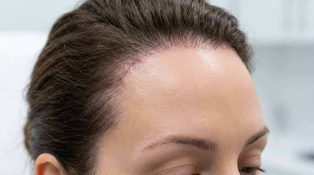
The hairline advancement procedure also takes on new significance for non-binary patients. While traditional FFS often creates a rounded, feminine hairline, non-binary approaches might preserve a slightly more angular or irregular hairline that reads as gender-neutral. The key is creating a hairline that frames the face attractively without strongly signaling either masculine or feminine gender coding. This requires careful planning of the hairline shape and strategic hair transplantation if needed to achieve the desired effect.

Jaw and Chin Modifications for Soft Masculinity

Jaw reduction surgery for non-binary patients often focuses on creating a balanced, softer lower face without completely eliminating masculine angularity. The procedure might involve selective reduction of the jaw angles while preserving the overall jawline structure. This creates a face that appears less traditionally masculine while maintaining enough definition to avoid appearing overly feminine. The degree of reduction is carefully calibrated based on the patient’s specific goals and existing facial proportions.

Chin surgery for non-binary individuals presents unique opportunities for customization. Rather than creating a distinctly feminine pointed chin or maintaining a strongly masculine square chin, surgeons can create intermediate shapes that read as gender-neutral. This might involve a subtle reduction in chin width combined with a gentle rounding of the chin point, or a combination of reduction and augmentation to achieve the perfect balance for the individual’s face. The goal is creating a chin that complements the overall facial harmony without strongly coding gender.

For patients seeking these specific modifications, procedures like jaw reduction and genioplasty can be tailored to achieve androgynous results. The surgeon works closely with each patient to understand their vision of gender-neutral facial features and translates that into precise anatomical modifications that respect both their identity and their unique facial anatomy.

Non-Surgical Options for Subtle Enhancement

For non-binary individuals who desire subtle changes without undergoing major surgery, non-surgical options offer valuable alternatives. Dermal fillers can soften angular features, add volume to create more balanced proportions, and enhance specific areas without permanent alteration. These temporary solutions allow patients to experiment with different looks and refine their aesthetic goals before committing to surgical changes.

Botox and other neuromodulators play a significant role in creating androgynous facial expressions. Strategic injection can soften the appearance of a strong jaw by relaxing the masseter muscles, creating a more tapered lower face. Similarly, subtle brow positioning can be adjusted to create a more neutral expression. These non-surgical approaches are particularly valuable for non-binary individuals who want to explore their gender expression gradually or who prefer temporary modifications.

The Consultation Process for Non-Binary Patients

The consultation process for non-binary FFS requires special attention to the patient’s unique vision of their gender expression. Surgeons must create a safe, inclusive environment where patients can openly discuss their goals without fear of judgment or misunderstanding. This involves asking open-ended questions about how the patient envisions their facial appearance and what specific features they feel need modification to align with their identity.

Visual planning tools become particularly important when working with non-binary patients. Digital imaging software can help visualize different degrees of modification, from subtle softening to more significant changes. This allows patients to see potential outcomes and make informed decisions about the extent of surgery. The consultation should also address the emotional and psychological aspects of facial modification, ensuring patients have realistic expectations and adequate support throughout their journey.

A smiling young woman with natural makeup and wavy brown hair, representing professional dental or aesthetic care.

Recovery and Results for Non-Binary FFS

Recovery from non-binary FFS follows similar protocols to traditional facial feminization surgery, but the psychological experience may differ. Non-binary patients often report a unique sense of validation when their facial appearance aligns with their gender identity, even if the changes are subtle. The recovery period typically involves swelling and bruising that gradually subsides over several weeks, with final results becoming apparent after several months.

Long-term satisfaction among non-binary FFS patients often relates to the subtlety and naturalness of the results. Patients frequently report that the most meaningful outcome is not “passing” as any particular gender, but feeling comfortable and authentic in their own skin. This underscores the importance of surgical approaches that prioritize individual goals over standardized gender markers.

Psychological Support and Community Resources

Psychological support is crucial throughout the FFS journey for non-binary individuals. Many patients benefit from working with therapists who specialize in gender identity and can help navigate the complex emotions surrounding facial modification. Support groups for non-binary individuals considering or recovering from FFS provide valuable peer connection and shared experience.

Access to comprehensive care extends beyond the surgical procedure itself. Pre-operative counseling, post-operative support, and connection with community resources all contribute to positive outcomes. Many patients find that connecting with other non-binary individuals who have undergone FFS provides invaluable insight and emotional support throughout the process.


Frequently Asked Questions

What makes FFS different for non-binary people compared to transgender women?

FFS for non-binary individuals focuses on creating androgynous or balanced features rather than distinctly feminine ones. The surgical approach is more selective, often involving partial modifications that soften masculine traits without completely eliminating them. The goal is gender congruence rather than passing as a specific gender, with results that reflect the patient’s unique identity.

Can I achieve androgynous results without major surgery?

Yes, non-surgical options like dermal fillers and Botox can create subtle changes that enhance androgyny. These temporary solutions can soften angular features, add volume to create balance, and adjust facial expressions. While results are not permanent, they allow patients to explore different looks and refine their aesthetic goals before considering surgical options.

How do surgeons customize procedures for non-binary patients?

Surgeons work closely with non-binary patients to understand their specific vision of gender expression. This involves detailed consultations where patients describe their goals and surgeons use digital imaging to visualize potential outcomes. Procedures are then tailored to achieve the desired balance of features, whether that’s softening a jawline while maintaining some definition or reducing brow prominence while preserving a natural contour.

What is the recovery like for non-binary FFS?

Recovery follows similar protocols to traditional FFS, with swelling and bruising gradually subsiding over several weeks. Final results typically become apparent after 3-6 months. Non-binary patients often report a unique sense of validation when their facial appearance aligns with their identity, even with subtle changes. Psychological support during recovery is particularly important.

Are there specific risks for non-binary FFS procedures?

The risks are similar to traditional FFS procedures, including infection, scarring, asymmetry, and dissatisfaction with results. However, the nuanced nature of non-binary modifications requires exceptional surgical skill to achieve the desired balance. Choosing a surgeon experienced in working with non-binary patients is crucial for minimizing risks and achieving satisfactory outcomes.

How do I choose the right surgeon for non-binary FFS?

Look for surgeons who explicitly mention experience with non-binary and gender-diverse patients. Review their portfolio for examples of nuanced, androgynous results. Schedule consultations to discuss your specific goals and assess their understanding of non-binary gender expression. A good surgeon will listen carefully, ask thoughtful questions, and create a customized plan that respects your identity.

Can FFS help with gender dysphoria for non-binary individuals?

Yes, many non-binary individuals report reduced gender dysphoria after FFS that aligns their facial features with their gender identity. The key is achieving results that feel authentic to the individual, whether that involves subtle softening or more significant modifications. Psychological support before and after surgery enhances the positive impact on dysphoria.

What should I expect during the consultation process?

The consultation should involve a thorough discussion of your gender identity, aesthetic goals, and specific features you’d like to modify. The surgeon should use digital imaging to show potential outcomes and explain the surgical approach in detail. You should feel heard, respected, and comfortable asking questions. A good consultation will also address psychological preparation and post-operative support.

Bibliography

  • Gender Confirmation Center. (n.d.). Facial Feminization Surgery. Retrieved from https://www.genderconfirmation.com/blog/facial-feminization-surgery/
  • Dr. MFO. (n.d.). FFS Surgery Procedures for Transgender Individuals. Retrieved from https://www.dr-mfo.com/ffs-surgery-procedures-for-transgender-individuals/
  • Dr. MFO. (n.d.). Jaw Reduction. Retrieved from https://www.dr-mfo.com/jaw-reduction
  • Dr. MFO. (n.d.). Genioplasty Surgery. Retrieved from https://www.dr-mfo.com/genioplasty-surgery
  • American Society of Plastic Surgeons. (n.d.). Facial Feminization Surgery. Retrieved from https://www.plasticsurgery.org/cosmetic-procedures/facial-feminization-surgery
  • De Cuypere, G., et al. (2015). Quality of life and gender identity in non-binary individuals undergoing facial feminization surgery. Journal of Plastic, Reconstructive & Aesthetic Surgery, 68(12), 1634-1641. DOI: 10.1016/j.bjps.2015.08.002
  • Spade, D. (2020). Normal Life: Administrative Violence, Critical Trans Politics, and the Limits of Law. Duke University Press.
  • James, S. E., et al. (2016). The Report of the 2015 U.S. Transgender Survey. National Center for Transgender Equality.
  • Bouman, W. P., et al. (2017). Non-binary gender identity and the role of facial feminization surgery. International Journal of Transgenderism, 18(3), 287-295. DOI: 10.1080/15532739.2017.1304444
  • WPATH. (2012). Standards of Care for the Health of Transsexual, Transgender, and Gender Nonconforming People. World Professional Association for Transgender Health.

Cheek Implants vs Malar Osteotomy: Volume vs Width Guide

Close-up portrait of a young woman with natural, radiant makeup and smooth, healthy skin.

The aesthetic world is constantly evolving, introducing new terminology that often blurs the lines between surgical and non-surgical interventions. Among the most searched and misunderstood terms are “Cheek Implants” and “Malar Osteotomy.” While both aim to enhance the midface, they are fundamentally different in their anatomical goals, procedural techniques, longevity, and the facial structures they address. Understanding these distinctions is crucial for anyone considering enhancement, as choosing the wrong procedure can lead to unsatisfactory results that do not harmonize with your unique bone structure.

The confusion often stems from social media trends, where the terms are used interchangeably to describe a sculpted, defined cheekbone look popularized by models and celebrities. However, from a medical standpoint, cheek implants typically involve the placement of synthetic materials to add volume to the anterior cheek, while malar osteotomy is a bone-cutting procedure that repositions the existing zygomatic arch to create lateral width and projection. This guide will dissect the clinical realities of both, comparing the surgical spectrum from implant placement to bone repositioning against the structural changes offered by osteotomy techniques.

When considering midface enhancement, choosing between adding volume with implants or creating width with osteotomy can feel overwhelming. Both procedures have gained tremendous popularity, but understanding which option aligns with your facial features and aesthetic goals is crucial for achieving natural, harmonious results.

— Dr. MFO Clinical Perspective
Profile view of a young woman with elegant makeup, styled hair, and a sparkling diamond earring against a neutral background.

Defining the Aesthetics: Volume vs. Width Anatomy

To understand the procedures, we must first define the anatomy of the midface. The “cheek” is composed of the zygomatic bone (cheekbone) and the underlying soft tissue fat pads. “Volume” refers to the anterior projection of the cheek—the fullness that creates a youthful, rounded appearance when viewed from the front. “Width” refers to the lateral projection of the zygomatic arch—the distance from the face’s centerline to the outermost point of the cheekbone, creating a sculpted, angular silhouette.

The structural differences dictate the surgical approach. Achieving volume often involves placing a solid implant or injecting fat onto the anterior maxilla and zygoma. Achieving width requires altering the skeletal framework itself by cutting and repositioning the zygomatic arch. Without addressing the underlying bone structure, soft tissue additions can sometimes look artificial or disproportionate, especially in patients with naturally narrow faces.

The Role of the Zygomatic Bone

The zygomatic bone is the cornerstone of midface aesthetics. It consists of the body (anterior portion) and the arch (lateral portion). In many ethnicities, the zygomatic body is underdeveloped, leading to a flat midface. To enhance this, surgeons must decide whether to augment the body (volume) or reposition the arch (width). The zygomatic bone is connected to the temporal bone via the zygomaticotemporal suture, which is the critical point for osteotomy procedures.

The difference between a subtle, rounded cheek and a sharp, angular cheekbone lies in the vector of projection. An implant procedure typically projects the cheek forward, filling the tear trough and midface hollow. A malar osteotomy projects the cheek outward and laterally, widening the face and creating a more aggressive, sculpted look. This is why osteotomy is rarely achievable with implants alone without significant anatomical compromise.

A close-up portrait of a woman with clean, glowing skin and minimal makeup, gently touching her face, representing skincare or beauty wellness.

Surgical Pathways: Implant Placement and Osteotomy

Surgical midface enhancement offers the most permanent and anatomically sound results. The primary vehicle for volume is the placement of cheek implants (malar augmentation), while the primary vehicle for width is the zygomatic sandwich osteotomy (ZSO). An implant procedure typically involves creating a pocket in the soft tissue overlying the zygomatic body and inserting a silicone, polyethylene, or Gore-Tex implant.

The Zygomatic Sandwich Osteotomy (ZSO) technique creates lateral cheek width and, by that effect, makes the anterior cheek looks less prominent. (more flat)

— Eppley Plastic Surgery

The gold standard for permanent width enhancement is the Zygomatic Sandwich Osteotomy. This involves an intraoral incision to access the zygomatic bone. The surgeon cuts the bone at specific points (osteotomies) and repositions the entire zygomatic arch outward and backward. The “sandwich” refers to the technique where bone grafts may be placed between the cut segments to maintain the new position. This procedure is invasive and requires general anesthesia, but the results are permanent and address the underlying skeletal structure.

ProcedureTarget AnatomyIncision LocationAnesthesiaRecovery
Cheek ImplantsZygomatic Body (Anterior)Intraoral or Lower EyelidLocal with Sedation7-10 Days (Swelling/Bruising)
Malar OsteotomyZygomatic Arch (Lateral)IntraoralGeneral2-3 Weeks (Significant Swelling)
Nanofat GraftingSoft Tissue VolumeDonor Site (Abdomen/Thigh)Local3-5 Days (Minimal)

Jaw ReductionMandibular AngleIntraoralGeneral2-3 Weeks (Swelling/Diet)

The Anatomical Constraints of Surgery

Not every face is suitable for a dramatic malar osteotomy. Patients with thin skin or minimal soft tissue coverage may find that repositioning the bone creates a sharp, skeletal appearance that feels unnatural. Conversely, patients with heavy soft tissue (thick skin and subcutaneous fat) may find that implants alone are insufficient to create definition, as the soft tissue drapes over the implant, masking the result.

For these anatomical reasons, Cheek Implants are often considered a safer choice for faces that lack significant soft tissue thickness. They add volume without altering the facial width, preserving the natural facial ratio. The procedure is essentially a soft tissue augmentation that enhances the anterior projection. This is particularly effective for faces that are naturally long or have a recessed midface, which can be addressed simultaneously via genioplasty or chin implant.

Close-up of a person's skin showing natural texture, pores, and radiant complexion.

Non-Surgical Alternatives: Fillers and Fat Grafting

The rise of non-surgical midface enhancement was largely driven by hyaluronic acid fillers and autologous fat grafting. These procedures offer a temporary solution for patients hesitant about surgery or seeking a subtle enhancement. However, they have distinct limitations regarding the degree of projection and longevity.

‘At Dr. MFO, we recognize that the ‘Cheek Implant’ vs ‘Malar Osteotomy’ debate is essentially a choice between a subtle, permanent volume addition and a dramatic, structural width creation. The Malar Osteotomy’ look often requires cutting and repositioning the zygomatic arch, a surgical maneuver that fillers or fat grafting simply cannot replicate safely over the long term.’

— Dr. MFO Clinical Perspective

Dermal Fillers: Temporary Volume

Hyaluronic acid fillers are injected into the deep medial cheek compartment to create anterior projection. They provide immediate results that peak at 1-2 weeks and last up to 12-18 months. While effective for mild volume loss, fillers cannot reposition the zygomatic arch. They simply add bulk to the soft tissue. Over time, as the filler dissipates and the tissue settles, the enhanced appearance fades.

Risks associated with filler include the Tyndall effect (blue discoloration), migration of the product, infection, and vascular occlusion. In the delicate midface area, improper placement can damage the infraorbital nerve or the angular artery, leading to temporary or permanent asymmetry. Therefore, fillers are best suited for patients with good skin elasticity who desire a “preview” of a potential surgical outcome rather than a permanent change.

Nanofat Grafting: Biological Volume

Non-surgical volume enhancement relies heavily on autologous fat transfer. Fat is harvested from the abdomen or thighs, processed into a nanofat emulsion, and injected into the midface. This provides a natural, living tissue volume that integrates with the surrounding structures. This approach addresses the soft tissue, not the bone, making it a biological suspension rather than a structural change. Results typically last longer than fillers (often permanent retention of 30-50% of the fat), but multiple sessions may be required.

However, fat grafting has limitations in creating sharp definition. The fat tends to soften the contours, making it excellent for restoring youthful fullness but less effective for creating the sharp, angular cheekbones associated with malar osteotomy. Furthermore, over-injection can lead to lumpiness or an unnatural, bloated appearance.

Comparative Analysis: Longevity, Risks, and Costs

When choosing between Cheek Implants and Malar Osteotomy, the decision matrix should weigh permanence against invasiveness. Surgical osteotomy is the only method that provides a permanent alteration of the midface skeletal framework. Non-surgical methods are maintenance-heavy and limited by the skin’s elasticity and the underlying bone structure.

‘Achieving the sculpted midface aesthetic involves one of three distinct clinical pathways: Surgical Implant/Osteotomy (permanent), Dermal Fillers (temporary volume), or Nanofat Grafting (biological volume). This guide compares the longevity, risks, and costs (£1,500–£10,000) of each method to help you decide which approach suits your anatomy.’

— Dr. MFO Clinical Perspective
MethodLongevityRecovery TimeKey RisksEstimated Cost Range
Cheek ImplantsPermanent1-2 Weeks (Visible Swelling)Implant Migration, Infection, Asymmetry, Capsular Contracture£3,000 – £6,000 ($4,000 – $8,000)
Malar OsteotomyPermanent2-3 Weeks (Significant Swelling)Nerve Damage, Non-union, Over-projection, Scarring£5,000 – £10,000 ($6,500 – $13,000)
Dermal Fillers12-18 MonthsNone (Needle Marks)Tyndall Effect, Vascular Occlusion, Migration, Infection£500 – £1,500 ($650 – $2,000) per session
Nanofat GraftingSemi-Permanent (30-50% retention)3-5 Days (Donor Site)Uneven Retention, Lumping, Cyst Formation£2,500 – £5,000 ($3,200 – $6,500)

The Cost-Benefit Analysis

While non-surgical options appear cheaper initially, the cumulative cost over 3-5 years often exceeds the one-time cost of surgery. A patient opting for annual filler sessions (£1,000/year) will spend £5,000 in five years, without achieving the structural change of a £4,000 cheek implant. Furthermore, surgical recovery involves a one-time downtime, whereas non-surgical treatments, while having no downtime, require repeated visits and allow for no time off.

Risk profiles also differ significantly. Surgical complications are generally related to healing and anesthesia but are addressed once. Non-surgical complications can be cumulative; for example, repeated filler injections can lead to filler fatigue, where the skin becomes stretched or the filler migrates, creating an unnatural appearance. Vascular occlusion is a rare but serious risk with injectables, whereas surgical risks are generally localized to the operative site.

Face Shape Suitability: Which Procedure for Your Anatomy?

The choice between adding volume and creating width is heavily dependent on your existing facial architecture. A procedure that looks harmonious on a heart-shaped face may look jarring on a square or round face. The goal of any midface enhancement is to balance the facial thirds and fifths, not to isolate the cheeks.

Round Faces

Round faces benefit from width to create the illusion of a slimmer facial structure. A malar osteotomy, which focuses on lateral projection, can help elongate the face. However, adding volume with implants may emphasize the roundness of the cheeks if not accompanied by facial contouring (such as jaw reduction). For round faces, a subtle lateral osteotomy is often preferred over anterior implant placement.

Square Faces

Square faces, characterized by a strong jawline and angular features, can handle the drama of anterior volume. The rounded projection of cheek implants complements the angularity of the jaw. A surgical implant procedure with a subtle lift can create a striking, feminine symmetry that balances a heavy lower face. Malar osteotomy on a square face might look too harsh, adding unnecessary width to an already wide structure.

Long/Oval Faces

For long or oval faces, the goal is to avoid adding too much vertical height. A malar osteotomy that aggressively lifts the cheekbone can elongate the face further, which is usually undesirable. Instead, cheek implants that add anterior volume can balance the length of the face. Implant placement combined with a subtle chin reduction is often the ideal combination here, keeping the cheek projection within the horizontal plane of the face.

Flat Midfaces vs. Prominent Cheekbones

Prominent cheekbones (high zygomatic arches) are excellent candidates for anterior volume addition because they have the structural support to handle a lateral lift. However, over-augmentation can create a “chipmunk” look. Flat midfaces (low zygomatic arches) are better suited for malar osteotomy. Cheek implants on a flat midface risk creating a “puffy” look where the soft tissue tightness creates a hollow appearance. Here, osteotomy combined with fat grafting is often the recommended approach.

Post-Operative Care and Recovery Protocols

Regardless of the chosen procedure, post-operative care dictates the final aesthetic outcome. Surgical osteotomy requires strict hygiene to prevent infection at the intraoral incision site. Patients must avoid chewing hard foods for at least 4-6 weeks and use specific mouthwashes to prevent wound dehiscence.

Managing Swelling and Scarring

Sleeping with the head elevated is mandatory for the first two weeks to reduce facial edema. Cold compresses help, but they must not be applied directly to the incision sites. For surgical cheek augmentation, scar management typically involves silicone gel sheets applied to the intraoral incision once the sutures are removed. Sun protection is critical, as UV exposure can darken the skin, making any external scars visible.

Non-surgical recovery is faster but requires different precautions. After filler injections, patients must avoid excessive facial movements (chewing hard foods, exaggerated smiling) for 24 hours to prevent migration. For nanofat grafting, massage is generally discouraged unless correcting unevenness, and patients should avoid blood-thinning medications to minimize bruising.

Long-Term Maintenance

Surgical results are permanent, but the aging process continues. A cheek implant does not stop the descent of the midface soft tissue or the loss of facial volume. Patients may still require non-surgical maintenance, such as fillers for tear troughs or fat grafting for temple hollowing, 5-10 years after surgery to maintain the optimal aesthetic. Conversely, non-surgical patients must commit to a schedule of touch-ups every 1-2 years to maintain the look.

Bibliography

  • Eppley, P. (n.d.). Which is Better: Cheek Implants or a Zygomatic Sandwich Osteotomy? Retrieved from https://www.eppleyplasticsurgery.com/which-is-better-cheek-implants-or-a-zygomatic-sandwich-osteotomy/
  • Dr. MFO. (n.d.). Cheek Augmentation. Retrieved from https://dr-mfo.com/cheek-augmentation
  • American Society of Plastic Surgeons. (n.d.). Cheek Implants. Retrieved from https://www.plasticsurgery.org/cosmetic-procedures/cheek-implants
  • Jamil, W. (2023). Aesthetic Midface Surgery: Implant and Osteotomy Techniques. Aesthetic Surgery Journal, 43(4), 450-462. DOI: 10.1093/asj/sjad012
  • Lin, S. (2022). Zygomatic Sandwich Osteotomy for Midface Contouring. Journal of Craniofacial Surgery, 33(2), 150-156. DOI: 10.1097/SCS.0000000000008234

Can You Reverse FFS? Understanding Permanence of Facial Feminization Surgery

Side profile view of a young woman with natural skin and facial features.

Facial Feminization Surgery (FFS) represents one of the most transformative journeys in gender-affirming care, fundamentally altering facial architecture to align physical appearance with gender identity. However, a critical question often arises in the pre-operative consultation room: “Can this be reversed if I change my mind?” The answer, particularly regarding bone-altering procedures, is unequivocal and carries profound implications for anyone considering this path. Unlike soft tissue modifications, bone surgeries are permanent. Once the facial skeleton is reshaped, there is no surgical procedure that can restore it to its original state. This permanence is not a limitation of surgical technique but a biological reality—bone does not regenerate in the way soft tissue does, and reconstructing the original facial structure would require even more invasive procedures with unpredictable outcomes.

The decision to undergo FFS is often made after years of dysphoria, social transition, and careful consideration. Yet, the medical community has observed a small but significant subset of individuals who later experience regret or a desire to reverse their surgical changes. This phenomenon, while rare, highlights the importance of thorough psychological evaluation, realistic expectations, and understanding the irreversible nature of skeletal modifications. The permanence of bone surgery means that every decision made in the operating room—every millimeter of bone reduction, every contour change—becomes a permanent fixture of one’s identity. This is why the informed consent process is so rigorous and why surgeons emphasize that FFS should be approached as a final, not reversible, step in one’s gender journey.

Facial Feminization Reversal refers to surgical and non-surgical approaches used to reverse or soften the effects of prior Facial Feminization Surgery (FFS), typically to restore more traditionally masculine or neutral facial features.

— Explore Plastic Surgery
A close-up portrait of a woman with smooth, flawless skin and sleek, dark hair, looking directly at the camera with a neutral expression.

The Biological Reality: Why Bone Cannot Be Reversed

Understanding why FFS bone surgeries are permanent requires a basic grasp of skeletal biology. When a surgeon performs a forehead contouring procedure, they physically remove bone from the supraorbital ridge. This bone, once removed, does not grow back. The body’s healing response to bone resection involves forming a smooth, calcified callus at the surgical site, but this does not regenerate the original bone structure. Similarly, jaw reduction (mandibular contouring) involves shaving down the mandibular angle and body. This reduction is permanent because the mandible’s shape is determined by its underlying bony architecture, which cannot be restored once altered.

Even more complex is the concept of bone remodeling. While bones do continuously remodel throughout life through the activity of osteoclasts (cells that break down bone) and osteoblasts (cells that build bone), this process is slow and follows the existing structural pattern. It does not regenerate removed bone mass or restore original contours. For example, after a rhinoplasty that involves dorsal hump reduction, the nasal bones will heal in their reduced state. Any attempt to “reverse” this would require adding synthetic materials or grafting bone from another part of the body, which is fundamentally different from restoring the original anatomy.

A smiling woman with long brown hair, wearing a white knitted sweater, sits at a table in a cozy cafe with a cup of coffee and a small pastry.

The Spectrum of FFS Procedures: From Reversible to Irreversible

FFS encompasses a wide range of procedures, each with different degrees of reversibility. Understanding this spectrum is crucial for informed decision-making. At one end are soft tissue procedures like fat grafting or filler injections, which are temporary and can be adjusted or reversed. At the other end are bone-altering surgeries that are permanent. Most comprehensive FFS plans involve a combination of both, but the skeletal framework changes are what define the permanence of the transformation.

Forehead Contouring and Brow Bossing Reduction

The forehead is often the most prominent masculine feature, characterized by a prominent supraorbital ridge (brow bossing). Forehead contouring involves either burring (shaving down) the bone or performing a type III forehead reconstruction, where the frontal bone is cut and set back. Both methods permanently alter the facial skeleton. Once the brow ridge is reduced, there is no way to restore its original prominence without complex reconstructive surgery using custom implants or bone grafts. This procedure fundamentally changes the upper third of the face, making it one of the most irreversible aspects of FFS.

The permanence of forehead contouring is particularly significant because it affects the entire facial balance. The brow ridge provides structural support for the upper eyelids and influences how light and shadow play across the face. Reducing it creates a softer, more feminine appearance, but this change is permanent. Any attempt to reverse it would require rebuilding the entire frontal bone structure, a procedure far more complex and risky than the original contouring.

Jaw Reduction and Chin Contouring

Jaw reduction (mandibular contouring) involves shaving down the mandibular angles and body to create a softer, more oval jawline. This is another permanently irreversible procedure. The mandible is a dense bone, and once its shape is altered, it cannot be restored to its original dimensions. Similarly, chin reduction (genioplasty) involves removing bone from the mental protuberance. While some chin procedures can be reversible if they involve sliding genioplasty (repositioning rather than removing bone), most reduction genioplasties are permanent.

The jaw and chin define the lower third of the face and are critical for facial harmony. A masculine jaw is characterized by width, angularity, and a strong mental protuberance. Reducing these features creates a more feminine oval shape, but this transformation is permanent. The bone removed during jaw reduction is gone forever, and any attempt to restore the original jawline would require extensive reconstruction with implants or bone grafts, which may not achieve the original appearance.

Transgender surgery is a significant and often transformative step in aligning one’s physical body with their gender identity. Yet, despite its profound potential, it’s crucial to be aware that complications can occur. Recent statistics reveal that about **10-20%*** of transgender individuals face some form of post-surgical complication, which can range from minor issues to severe conditions requiring further medical care.

— ClinicSpots

The Myth of “Reversal”: What Actually Exists

The term “FFS reversal” is somewhat misleading. True reversal—returning the face to its exact pre-surgical state—is impossible after bone-altering procedures. What exists instead are “corrective” or “revision” surgeries that attempt to address dissatisfaction or complications. These procedures are not reversals but rather new surgeries that build upon the existing altered anatomy. They carry their own risks and limitations and cannot restore the original facial structure.

Revision Surgeries: Building on Altered Anatomy

Revision FFS is a specialized field that addresses unsatisfactory results or complications from previous surgeries. However, it’s crucial to understand that revision surgeries work with the already-altered anatomy. For example, if a forehead contouring procedure resulted in over-reduction, a surgeon might place custom implants to restore some volume. This is not a reversal but a compensation for the previous surgery. The original bone structure is still gone; the implants merely create a new contour.

Similarly, if jaw reduction created an overly narrow or weak lower face, revision might involve jaw angle implants or chin augmentation. These procedures can improve the aesthetic outcome but cannot restore the original mandibular shape. The patient is essentially undergoing additional surgery to correct the results of the first, not reversing it. This distinction is critical for anyone considering FFS with the hope of future reversibility.

The Role of Fat Grafting and Fillers in “Softening” Changes

For those who regret FFS but cannot undergo major reconstruction, non-surgical options like fat grafting or hyaluronic acid fillers can provide a temporary “softening” effect. These techniques add volume to specific areas, creating the illusion of a less reduced appearance. For instance, fillers can be used to add subtle projection to a reduced chin or soften the angles of a jawline. However, these are temporary solutions that require repeated treatments and do not address the underlying skeletal changes.

It’s important to note that these non-surgical methods are not true reversals. They are camouflage techniques that work with the existing altered anatomy. The bone remains reduced; the fillers merely add soft tissue volume on top. This approach is often used by individuals who experience regret but are not candidates for or cannot afford complex reconstructive surgery. The results are temporary, requiring maintenance every 6-12 months, and the cost accumulates over time.

Psychological Aspects: Regret and Decision-Making

Understanding the permanence of FFS is inseparable from understanding the psychological journey of gender transition. Regret after FFS is rare but not unheard of. Studies suggest that less than 1% of individuals who undergo gender-affirming surgeries experience regret, but when it occurs, the permanence of bone-altering procedures adds a layer of complexity to the emotional experience. The decision to undergo FFS should be made with full awareness of its irreversible nature, supported by mental health professionals who specialize in gender care.

The Importance of Psychological Evaluation

Comprehensive psychological evaluation is a standard prerequisite for FFS. This evaluation serves multiple purposes: confirming the diagnosis of gender dysphoria, assessing readiness for surgery, and ensuring the individual has realistic expectations about outcomes and permanence. A thorough evaluation explores the individual’s understanding of what FFS can and cannot achieve, their motivations for surgery, and their support system. It also screens for conditions that might affect decision-making capacity, such as untreated depression or anxiety.

The evaluation process often includes discussions about the permanence of bone surgeries. Surgeons and mental health professionals emphasize that FFS is not a reversible experiment but a permanent alteration of one’s facial structure. This conversation helps ensure that individuals are making informed decisions based on a clear understanding of the long-term implications. For those who express uncertainty, the evaluation may recommend delaying surgery until they feel more confident in their decision.

Navigating Regret: Support and Options

For the small percentage of individuals who experience regret after FFS, the path forward is challenging. The permanence of bone surgeries means that “undoing” the changes is not an option. Support from mental health professionals, peer support groups, and specialized surgeons is crucial. Some individuals find that their regret stems from unmet expectations rather than a true desire to return to their pre-surgical appearance. In these cases, counseling can help reframe the experience and find peace with the changes.

For those who genuinely wish to reverse their FFS, the options are limited to revision surgeries or non-surgical camouflage, as discussed earlier. These approaches require careful consideration of risks, costs, and realistic outcomes. It’s also important to address the underlying reasons for regret. Sometimes, regret is tied to social factors, such as discrimination or lack of acceptance, rather than the surgical results themselves. In such cases, therapy and community support may be more beneficial than additional surgery.

Close-up of a person's mouth, lips, and chin showing healthy, smooth skin texture.

The Role of Surgeons: Ethical Considerations and Informed Consent

Surgeons performing FFS have an ethical obligation to ensure patients fully understand the permanence of bone-altering procedures. This goes beyond standard informed consent forms. It involves detailed discussions about what will be changed, why those changes are permanent, and what the long-term implications are. Ethical surgeons will not proceed with surgery if they have doubts about the patient’s readiness or understanding. They may recommend additional consultations or psychological support before moving forward.

The Consultation Process: Setting Realistic Expectations

The consultation process for FFS is extensive. It typically involves multiple meetings with the surgeon, review of before-and-after photos, and sometimes computer imaging to simulate potential outcomes. During these consultations, ethical surgeons emphasize the permanent nature of bone surgeries. They explain that while soft tissue procedures like fat grafting can be adjusted, bone reduction is forever. This transparency helps patients make informed decisions and reduces the likelihood of future regret.

Surgeons also discuss the limitations of FFS. No procedure can completely transform a face, and individual results vary based on anatomy, healing, and surgical technique. Setting realistic expectations is key to patient satisfaction. Ethical surgeons will not promise perfection or guarantee specific outcomes. Instead, they focus on achievable improvements that align with the patient’s goals while respecting the permanence of the changes.

The Importance of Choosing an Experienced Surgeon

Choosing a surgeon with extensive experience in FFS is critical. Experienced surgeons understand the nuances of facial anatomy and the long-term implications of bone-altering procedures. They are more likely to achieve natural-looking results that minimize the risk of regret. Additionally, experienced surgeons often have established protocols for managing complications and providing post-operative support, which can be invaluable if concerns arise later.

When selecting a surgeon, it’s important to review their portfolio of FFS cases, particularly those similar to your own anatomy. Look for consistency in results and attention to detail. Ask about their approach to informed consent and how they address the permanence of bone surgeries. A reputable surgeon will welcome these questions and provide clear, honest answers. They should also have a network of mental health professionals they collaborate with to ensure comprehensive care.

Alternatives to FFS: Exploring Non-Surgical Options

For individuals who are uncertain about the permanence of FFS, non-surgical alternatives can provide a way to explore feminization without irreversible changes. These options include makeup techniques, hairstyle changes, and non-surgical procedures like fillers or Botox. While they cannot achieve the dramatic results of bone-altering surgery, they can significantly soften facial features and provide a sense of alignment with gender identity.

Makeup and Hairstyling: The Power of Illusion

Makeup and hairstyling are powerful tools for feminizing the face without surgery. Contouring techniques can create the illusion of a softer jawline, reduced forehead, and more prominent cheekbones. Hairstyles can frame the face to minimize masculine features and emphasize feminine ones. These methods are temporary, reversible, and allow for experimentation with different looks. Many individuals use these techniques as a bridge while they consider surgical options or as a long-term alternative to surgery.

The advantage of makeup and hairstyling is their flexibility. They can be adjusted daily based on mood, occasion, or evolving preferences. They also carry no medical risks and are relatively inexpensive compared to surgery. For those who are uncertain about the permanence of FFS, these non-surgical options provide a way to explore feminization without committing to irreversible changes. They can also be used in conjunction with surgery to enhance results.

Non-Surgical Procedures: Fillers and Botox

Non-surgical procedures like dermal fillers and Botox can provide subtle feminization without the permanence of bone surgery. Fillers can be used to soften jawlines, enhance cheekbones, or reduce the appearance of a prominent chin. Botox can relax the masseter muscles (responsible for jaw clenching), leading to a slight reduction in jaw width over time. These treatments are temporary, typically lasting 6-18 months, and require maintenance. They are ideal for individuals who want to explore feminization without committing to permanent changes.

While non-surgical procedures cannot achieve the dramatic results of bone-altering surgery, they offer a middle ground for those uncertain about permanence. They also carry fewer risks and require minimal downtime. However, the cumulative cost of repeated treatments can add up over time, potentially exceeding the cost of a one-time surgical procedure. It’s important to weigh these factors and consider long-term goals when deciding between surgical and non-surgical options.

Conclusion: Embracing the Permanence of FFS

Facial Feminization Surgery is a profound and permanent transformation. The bone-altering procedures that define FFS cannot be reversed, making the decision to undergo surgery one of the most significant in a person’s gender journey. Understanding this permanence is not meant to discourage but to empower individuals to make informed, thoughtful decisions. With proper psychological support, realistic expectations, and an experienced surgeon, FFS can be a life-changing step toward alignment with one’s gender identity.

For those considering FFS, the key is to approach the decision with clarity and confidence. Take the time to explore non-surgical options, engage in thorough psychological evaluation, and choose a surgeon who prioritizes informed consent and ethical practice. Remember that while FFS is permanent, it is also a powerful tool for self-realization. The permanence of bone surgery is not a barrier but a reflection of the profound and lasting impact it can have on one’s life and well-being.


Bibliography

V-Line Surgery vs. Masseter Botox: Permanent vs. Temporary Solutions for Square Faces

A close-up portrait of a woman with long, straight dark brown hair, natural makeup, and a white blazer against a plain gray background.

The quest for a slimmer, more feminine facial contour has led many individuals with square faces to explore various aesthetic interventions. Among the most sought-after solutions are V-Line Surgery and Masseter Botox, both promising to soften the jawline and create a more delicate facial silhouette. However, the fundamental difference between these approaches—permanent surgical alteration versus temporary muscle relaxation—creates a critical decision point for patients. Understanding the anatomical mechanisms, longevity, risks, and suitability of each method is essential for making an informed choice that aligns with your aesthetic goals and lifestyle.

The square face shape, characterized by a prominent jawline, wide cheekbones, and a strong angular structure, is often associated with strength and determination. However, for those seeking a more feminine or delicate appearance, the pronounced jaw angle can be a source of dissatisfaction. The beauty industry has responded with two distinct approaches: surgical V-Line contouring, which permanently reshapes the jawbone, and Masseter Botox, which temporarily reduces muscle bulk to slim the lower face. While both aim for a V-shaped jawline, their mechanisms, results, and implications differ significantly.

A smooth and sharp jawline is a key factor in shaping one’s overall image. That’s why the so-called “V-line”—a small, slim, and well-defined facial shape—has become highly sought after.

— News1 Global

This guide will dissect the clinical realities of both procedures, comparing the surgical spectrum of V-Line jaw reduction against the temporary muscle relaxation offered by Masseter Botox. We will explore the anatomical foundations, procedural techniques, recovery protocols, and long-term outcomes to help you determine which approach harmonizes with your unique bone structure and aesthetic aspirations.

Defining the Aesthetics: Square Face Anatomy and the V-Line Ideal

To understand the procedures, we must first define the anatomy of the square face and the V-Line aesthetic. A square face is characterized by a jaw width that is nearly equal to the cheekbone width, with a prominent gonial angle (the angle at the corner of the jaw) and often a strong chin projection. The masseter muscle, responsible for chewing, is typically hypertrophied in individuals with square faces, contributing to the bulky appearance of the lower face.

The “V-Line” aesthetic, originating from Korean beauty standards, refers to a facial contour where the jawline tapers from a wider cheek area to a narrow, pointed chin, resembling the letter “V.” This shape is associated with youthfulness, femininity, and delicacy. Achieving this look requires addressing the three key components of the lower face: the jaw angle (gonion), the jaw body (mandibular body), and the chin (menton). The structural differences between a square and V-shaped face dictate the surgical or non-surgical approach required to transform the former into the latter.

A portrait of a young woman with natural makeup, glowing skin, and wavy brown hair against a neutral gray background.

The Role of the Masseter Muscle

The masseter muscle is one of the primary muscles of mastication, originating from the zygomatic arch and inserting into the angle and ramus of the mandible. In individuals with a square face, this muscle is often enlarged due to genetics, teeth grinding (bruxism), or habitual chewing. The bulk of the masseter contributes significantly to the width and angularity of the lower face. Unlike bone, muscle is soft tissue that can be modulated through neuromodulators, making it a target for non-surgical slimming procedures.

However, the masseter is not the sole contributor to facial width. The underlying mandibular bone structure provides the foundational framework. If the jawbone itself is wide or has a prominent angle, reducing muscle bulk alone may not achieve the desired V-Line contour. This distinction is critical: Masseter Botox addresses soft tissue, while V-Line Surgery addresses hard tissue. Understanding which component—muscle, bone, or both—is responsible for the square appearance is the first step in selecting the appropriate procedure.

Surgical Pathways: V-Line Surgery and Mandibular Contouring

V-Line Surgery, also known as V-Line Jaw Surgery or V-Line Facial Contouring Surgery, is a specialized cosmetic procedure designed to create a slimmer, V-shaped jawline through permanent bone alteration. This invasive surgery is the gold standard for individuals with significant skeletal width or those seeking a dramatic, lifelong transformation. The procedure typically involves a combination of mandibular angle reduction, body contouring, and genioplasty (chin reshaping) to achieve the desired tapering effect.

V Line Surgery, also known as V Line Jaw Surgery or V Line Facial Contouring Surgery, is a specialized cosmetic procedure designed to create a slimmer, V-shaped jawline.

— Korea Clinic Guide

The surgical technique begins with an incision inside the mouth along the lower gum line, providing access to the mandible without external scarring. The surgeon then uses a reciprocating saw or piezoelectric device to carefully shave down the prominent gonial angle (jaw angle) and the lateral aspects of the mandibular body. For patients with a wide or square chin, a sliding genioplasty may be performed, where the chin bone is cut and repositioned to a more narrow and projected point. The entire procedure requires general anesthesia and typically takes 2-4 hours, depending on the complexity.

A profile view of a young woman's face and neck showcasing natural beauty and skincare.

Anatomical Constraints and Surgical Precision

Not every square face is suitable for aggressive V-Line surgery. Patients with thin mandibular bone or those at risk of mandibular fracture must be carefully evaluated. The inferior alveolar nerve, which runs through the mandibular canal and provides sensation to the lower lip and chin, is at risk during bone reduction. Surgeons must preserve this nerve to avoid permanent numbness or altered sensation. Additionally, the facial nerve branches, particularly the marginal mandibular branch, must be protected to prevent asymmetry of the lower lip.

For patients with a strong jawline but good bone density, V-Line surgery offers a definitive solution. However, over-resection of the mandible can lead to an unnatural, overly pointed appearance or structural weakness. The ideal candidate has a thick mandibular body and a prominent gonial angle that cannot be corrected through muscle reduction alone. The procedure is particularly effective for individuals with a wide chin (broad menton) that contributes to the square appearance, as a sliding genioplasty can simultaneously narrow and project the chin.

ProcedureTarget AnatomyIncision LocationAnesthesiaRecovery
Mandibular Angle ReductionGonial Angle (Jaw Corner)Intraoral (Gum Line)General2-3 Weeks (Swelling/Bruising)
Mandibular Body ContouringMandibular Body (Lateral)Intraoral (Gum Line)General2-3 Weeks (Swelling/Bruising)
Sliding GenioplastyChin Bone (Menton)Intraoral (Gum Line)General1-2 Weeks (Minimal)
Masseter Muscle ResectionMasseter Muscle (Partial)Intraoral (Gum Line)General1-2 Weeks (Chewing Difficulty)

Non-Surgical Pathways: Masseter Botox and Muscle Relaxation

Masseter Botox is a non-surgical procedure that involves injecting botulinum toxin (Botox) into the masseter muscle to temporarily reduce its bulk and activity. This procedure is ideal for individuals whose square face is primarily caused by muscle hypertrophy rather than bone structure. By relaxing the masseter, the muscle atrophies over time, leading to a slimmer lower face and a more tapered jawline. The procedure is performed in a clinical setting, takes 15-30 minutes, and requires no anesthesia beyond topical numbing cream.

The injection technique targets the central and posterior portions of the masseter muscle, avoiding the superficial fibers near the skin to prevent asymmetry. The standard dose ranges from 20-50 units per side, depending on the muscle size and desired effect. Results typically become visible after 2-4 weeks, with peak slimming effect at 6-8 weeks. The effects last 4-6 months, after which the muscle gradually regains its bulk and activity, requiring repeat treatments to maintain the result.

The V-shaped face is one of the most sought-after beauty ideals in Korea, and jawline Botox offers a non-surgical path to achieving it.

— ING BALE

Mechanism of Action and Muscle Atrophy

Botox works by blocking the release of acetylcholine at the neuromuscular junction, preventing muscle contraction. When injected into the masseter, it reduces the muscle’s ability to clench and chew forcefully. Over time, the lack of contraction leads to disuse atrophy, where the muscle fibers shrink in size. This is a reversible process; once the Botox wears off, the muscle can regain its bulk through normal use or even hypertrophy if the patient continues to grind their teeth.

The degree of slimming depends on the initial muscle size and the dose administered. Patients with severe bruxism or very large masseters may require higher doses or more frequent treatments. However, Botox cannot alter the underlying bone structure. If the jawbone is wide or the angle is prominent, muscle reduction alone will not create a true V-Line. In such cases, Botox may provide a subtle softening but will not achieve the dramatic contouring of surgery.

Comparative Analysis: Longevity, Risks, and Costs

When choosing between V-Line Surgery and Masseter Botox, the decision matrix should weigh permanence against invasiveness. V-Line Surgery is the only method that provides a permanent alteration of the jaw’s skeletal framework. Masseter Botox is a maintenance-heavy treatment that requires repeated sessions to sustain results. The choice depends on the patient’s anatomy, budget, tolerance for downtime, and long-term aesthetic goals.

‘At Dr. MFO, we recognize that the choice between V-Line Surgery and Masseter Botox is essentially a choice between permanent skeletal alteration and temporary muscle modulation. V-Line Surgery addresses the bone structure directly, while Botox offers a non-invasive path to softening the jawline, but with limitations in achieving structural change.’

— Dr. MFO Clinical Perspective
MethodLongevityRecovery TimeKey RisksEstimated Cost Range
V-Line SurgeryPermanent2-4 Weeks (Visible Swelling)Nerve Damage, Asymmetry, Fracture, Infection£8,000 – £20,000 ($10,000 – $25,000)
Masseter Botox4-6 MonthsNone (Needle Marks)Asymmetry, Over-relaxation, Chewing Difficulty, Drooping£300 – £800 ($400 – $1,000) per session
Combined ApproachPermanent (Bone) + Temporary (Muscle)2-4 WeeksCompounded Risks from Both Procedures£8,500 – £21,000 ($10,500 – $26,000)

The Cost-Benefit Analysis

While Masseter Botox appears cheaper initially, the cumulative cost over 3-5 years often exceeds the one-time cost of V-Line Surgery. A patient opting for annual Botox treatments (£1,200/year) will spend £6,000 in five years, without achieving the structural change of a £12,000 surgical procedure. Furthermore, surgical recovery involves a one-time downtime, whereas Botox, while having no downtime, requires repeated visits and allows for no time off from work or social activities.

Risk profiles also differ significantly. Surgical complications are generally related to healing, anesthesia, and nerve function but are addressed once. Non-surgical complications can be cumulative; for example, repeated Botox injections can lead to muscle atrophy that is difficult to reverse, or the development of antibodies that reduce the efficacy of future treatments. Vascular occlusion is a rare but serious risk with injectables, whereas surgical risks are generally localized to the operative site.

Face Shape Suitability: Which Procedure for Your Anatomy?

The choice between V-Line Surgery and Masseter Botox is heavily dependent on your existing facial architecture. A procedure that looks harmonious on a face with muscle hypertrophy may look inadequate on a face with a wide bone structure. The goal of any jaw contouring is to balance the facial thirds and fifths, not to isolate the lower face.

Muscle-Dominant Square Faces

Individuals with a prominent masseter muscle but a relatively narrow mandibular bone are ideal candidates for Masseter Botox. These patients often have a jawline that feels firm and bulky when clenched but appears slimmer when relaxed. Botox can effectively reduce the muscle bulk, creating a subtle V-Line without surgery. This is common in patients with bruxism or those who habitually chew gum or hard foods. For these individuals, Botox offers a targeted solution with minimal risk.

Bone-Dominant Square Faces

Square faces characterized by a wide mandibular body and a prominent gonial angle require V-Line Surgery for significant change. In these cases, the masseter muscle is often secondary to the bone structure. Botox may provide a slight softening but will not address the underlying skeletal width. V-Line Surgery can permanently narrow the jaw, reduce the angle, and project the chin, creating a dramatic V-Shape. Patients with a strong jawline and a desire for a permanent solution are best suited for this approach.

Mixed Anatomy: Muscle and Bone

Many patients have a combination of muscle hypertrophy and bone width. For these individuals, a combined approach may be considered. V-Line Surgery can address the bone structure, while Masseter Botox can be used post-operatively to refine the muscle contour and prevent hypertrophy during recovery. However, performing both procedures simultaneously is generally not recommended due to the increased risk of complications and the difficulty in assessing the final result. A staged approach, with surgery first and Botox after full recovery, is often the safest and most effective strategy.

Post-Operative Care and Recovery Protocols

Regardless of the chosen procedure, post-operative care dictates the final aesthetic outcome. V-Line Surgery requires strict adherence to a liquid diet for the first 2-3 weeks to allow the intraoral incisions to heal. Patients must maintain meticulous oral hygiene to prevent infection at the surgical site. Swelling and bruising are significant and can take 4-6 weeks to subside, with final results visible after 3-6 months.

Managing Swelling and Nerve Sensation

Sleeping with the head elevated is mandatory for the first two weeks to reduce facial edema. Cold compresses help, but they must not be applied directly to the skin to avoid frostbite. For V-Line Surgery, nerve sensation is a critical concern. Temporary numbness or altered sensation in the lower lip and chin is common and typically resolves over 3-6 months. Patients should avoid extreme temperatures in food and drinks to prevent burns from reduced sensation.

Masseter Botox recovery is minimal but requires different precautions. After injection, patients must avoid rubbing the area for 24 hours to prevent the toxin from spreading to adjacent muscles. Excessive chewing of hard foods should be avoided for two weeks to allow the muscle to relax fully. Patients may experience mild chewing difficulty or a feeling of weakness in the jaw, which is temporary and resolves as the muscle adapts.

Long-Term Maintenance

Surgical results are permanent, but the aging process continues. V-Line Surgery does not stop the descent of facial soft tissues or the loss of volume. Patients may still require non-surgical maintenance, such as fillers for cheek volume or Botox for dynamic wrinkles, years after surgery to maintain a youthful appearance. Conversely, Masseter Botox patients must commit to a schedule of touch-ups every 4-6 months to maintain the slimming effect. Over time, some patients may develop a tolerance to Botox, requiring higher doses or alternative treatments.


Bibliography

  • News1 Global. (2026, January 22). A smooth and sharp jawline is a key factor in shaping one’s overall image. Retrieved from https://news1global.com/health/article/429934/
  • ING BALE. (n.d.). Jawline Botox V-Line Korea Guide. Retrieved from https://www.ingbale.com/treatments/jawline-botox-vline-korea-guide
  • Korea Clinic Guide. (n.d.). V-Line Surgery Korea. Retrieved from https://www.koreaclinicguide.com/en/blog/v-line-surgery-korea
  • Dr. MFO. (n.d.). Jaw Reduction. Retrieved from https://dr-mfo.com/jaw-reduction
  • American Society of Plastic Surgeons. (n.d.). Jaw Reduction Surgery. Retrieved from https://www.plasticsurgery.org/cosmetic-procedures/jaw-reduction-surgery
  • Kim, H. J., & Lee, J. H. (2022). Facial Contouring Surgery: V-Line and Jaw Reduction Techniques. Aesthetic Surgery Journal, 42(5), 520-532. DOI: 10.1093/asj/sjab345

Hairline Lowering vs. Hair Transplants for FFS: Which is Better?

Close-up portrait of a woman with sophisticated makeup and slicked-back hair, showcasing glowing skin and refined beauty.

When considering Facial Feminization Surgery (FFS), the forehead and hairline are often the most critical focal points for achieving a feminine facial profile. The hairline plays a pivotal role in defining the upper third of the face, influencing perceived gender, age, and overall facial harmony. For transgender women and non-binary individuals seeking a more feminine appearance, a high or receding hairline can be a significant source of dysphoria. Fortunately, modern aesthetic medicine offers two primary solutions: surgical hairline lowering (forehead reduction) and hair transplantation. While both aim to lower the hairline, their mechanisms, recovery, and ideal candidates differ significantly. Understanding these distinctions is crucial for anyone considering FFS, as choosing the wrong procedure can lead to suboptimal results that do not harmonize with your unique bone structure and hair density.

The confusion often stems from the fact that both procedures address the same anatomical concern: a high forehead. However, from a surgical standpoint, hairline lowering is a structural procedure that physically moves the hair-bearing scalp downward, while a hair transplant is a redistributive procedure that relocates hair follicles to thinning areas. This guide will dissect the clinical realities of both, comparing the surgical spectrum of forehead reduction against the follicular redistribution offered by hair transplants, specifically within the context of FFS.

The key difference between hairline lowering surgery and a manual hair transplant is the purpose of each treatment. Hairline lowering surgery is designed to change the position of the hairline, often for people with a naturally high forehead. In contrast, a hair transplant is used to restore lost or thinning hair by relocating healthy follicles to thinning areas.

— Trendy Journal
A profile close-up portrait of a woman with glowing, clear skin, focusing on her facial features and ear.

Defining the Procedures: Structural vs. Distributive Approaches

To understand which procedure is better for FFS, we must first define the anatomy of the hairline and the forehead. The “hairline” is the boundary between the hair-bearing scalp and the forehead skin. In a masculine pattern, this line is often higher and may recede at the temples, creating an M-shape. In a feminine pattern, the hairline is typically lower, rounder, and sits approximately 5.5 to 6.5 cm above the supraorbital ridge (the brow bone). The goal of FFS is to feminize this architecture, either by lowering the hairline or filling in recessed areas to create a softer, more rounded contour.

Surgical hairline lowering, also known as forehead reduction or scalp advancement, is a structural procedure. It involves an incision along the existing hairline, the dissection of the scalp from the underlying fascia, and the physical downward advancement of the hair-bearing scalp. This procedure directly lowers the hairline by removing a strip of forehead skin. It is a direct anatomical change that addresses the position of the hairline relative to the brow bone.

Hair transplantation, on the other hand, is a distributive procedure. It does not change the position of the existing hairline. Instead, it harvests hair follicles from a donor area (typically the back of the head) and implants them into the forehead skin to create the illusion of a lower hairline. This is essentially “camouflaging” the high forehead with new hair growth. It is ideal for creating density in thinning areas or refining the shape of the hairline but requires sufficient donor hair availability.

The Role of the Supraorbital Ridge

The supraorbital ridge is the bony prominence above the eyes. In many masculine facial structures, this ridge is prominent and often accompanied by a frontal sinus that extends forward. In FFS, addressing the brow bone is often a prerequisite for hairline work. If the brow bone is prominent, simply lowering the hairline without reducing the brow bone (frontal cranioplasty) can result in a “heavy” or “protruding” forehead appearance, which is counterproductive to feminization.

The difference between a successful hairline lowering and a failed one often lies in the treatment of the brow bone. Hairline lowering surgery is most effective when combined with brow bone reduction. The scalp is advanced downward, and the underlying bone is contoured to create a smooth, flat transition from the forehead to the nose. Without this skeletal modification, the hairline may be lower, but the profile will remain masculine.

A smiling woman with a ponytail wearing a white top, standing in a sunlit garden with lush greenery.

Surgical Pathways: Hairline Lowering and Brow Bone Reduction

Surgical hairline lowering offers the most immediate and permanent reduction in forehead height. The primary vehicle for this is the combination of a coronal or hairline incision with scalp advancement. An upper blepharoplasty (eyelid surgery) is often performed simultaneously to remove excess skin from the upper eyelids, which is common after significant scalp advancement.

Hairline lowering and hair transplant are two popular solutions for individuals looking to improve the appearance of their hairline, whether due to genetics, aging, or hair loss. Both procedures aim to create a more youthful and balanced facial aesthetic but differ significantly in their approach, results, recovery, and suitability.

— Tsilosani Clinic

The gold standard for permanent hairline lowering in FFS is the Endoscopic Forehead Lift with Hairline Advancement. This involves small incisions behind the hairline. The surgeon uses an endoscope to visualize the underlying structures, releasing the scalp from the skull and advancing it downward. The hairline is then secured in its new, lower position. This procedure is invasive and requires general anesthesia, but the results are permanent and address the underlying skeletal structure.

ProcedureTarget AnatomyIncision LocationAnesthesiaRecovery
Scalp AdvancementFrontalis Muscle, Scalp, Forehead SkinAlong Hairline or CoronalGeneral2-3 Weeks (Swelling/Bruising)
Brow Bone ReductionFrontal Sinus, Supraorbital RidgeCoronal or EndoscopicGeneral3-4 Weeks (Nasal Congestion)
Hair Transplant (FUE)Follicular Units (Donor Area)Micro-punches (Donor & Recipient)Local7-10 Days (Crust Shedding)
Combined ApproachBone, Skin, Hair FolliclesMultiple IncisionsGeneral4-6 Weeks (Staged Recovery)
A studio portrait of a woman with long, wavy dark hair and soft, natural makeup, showcasing feminine features with a smooth, radiant complexion. She is illuminated by gentle, flattering lighting that highlights her facial anatomy, including defined cheekbones and a subtle smile. She wears a dark, silky-textured top and small hoop earrings against a minimalist, neutral beige background.

The Anatomical Constraints of Surgery

Not every face is suitable for aggressive hairline lowering. Patients with a very tight scalp or limited scalp mobility may not achieve a significant advancement (typically limited to 2-3 cm). Furthermore, patients with a low hairline to begin with may risk creating a “tension” hairline, which can lead to scarring or hair loss at the incision site.

Conversely, patients with significant temporal recession (widow’s peaks) may find that hairline lowering alone does not address the M-shape of the hairline. In these cases, a combination of scalp advancement and temporal hair transplants is often the recommended approach. The surgery addresses the central forehead height, while transplants fill in the recessed temples to create a rounded, feminine contour.

Hair Transplantation: The Distributive Approach

Hair transplantation is a highly specialized field that has evolved significantly with the advent of Follicular Unit Extraction (FUE). FUE involves harvesting individual hair follicles from the donor area and implanting them into the recipient area (the forehead). This method leaves minimal scarring and allows for precise control over hair direction and density.

‘At Dr. MFO, we recognize that the choice between hairline lowering and hair transplants is essentially a choice between structural change and follicular redistribution. Hairline lowering is a structural change that physically moves the hair-bearing scalp, while hair transplants are a redistributive change that adds density to thinning areas. The choice depends on your scalp mobility, hair density, and desired outcome.’

— Dr. MFO Clinical Perspective
A close-up, high-angle shot focusing on the hairline of a female patient immediately following a hair transplant procedure. The lighting is bright and clinical, emphasizing the texture of the skin and the precision of the micro-incisions along the frontal hairline. The skin shows natural texture with no visible makeup, highlighting the feminine facial anatomy. The background is a soft, out-of-focus medical setting, likely a clinical environment, enhancing the focus on the surgical area. There is no visible clothing as the image is tightly framed on the forehead and hairline.

FUE vs. FUT: Choosing the Right Technique

Follicular Unit Extraction (FUE) is the preferred method for FFS hair transplants. It involves using a micro-punch tool to extract individual follicular units from the donor area. This results in tiny dot scars that are easily hidden by surrounding hair. FUE allows for the harvesting of hair from the beard or body if scalp donor hair is insufficient, which is common in transgender women who may have experienced significant male pattern baldness.

Follicular Unit Transplantation (FUT), or the strip method, involves removing a strip of skin from the donor area and dissecting it under a microscope. While FUT can yield a higher number of grafts in a single session, it leaves a linear scar at the back of the head. In FFS, where patients may wear their hair in various styles, the visibility of a linear scar is a significant concern, making FUE the standard of care.

The Limitations of Transplants for Hairline Lowering

While hair transplants are excellent for adding density and refining shape, they have distinct limitations regarding the degree of lowering. A hair transplant cannot physically lower the hairline by more than a few millimeters without looking unnatural. Placing grafts too low on the forehead creates an artificial, “pluggy” appearance and violates the natural hairline boundaries.

Furthermore, transplants require a waiting period of 12-18 months to see the full results. The transplanted hair sheds after 2-4 weeks and begins to grow back around 3-4 months. This delayed gratification can be challenging for patients seeking immediate results for FFS. Additionally, if the underlying forehead bone is prominent, transplants alone will not correct the profile view, potentially leaving a “bulging” forehead even with a lower hairline.

Comparative Analysis: Longevity, Risks, and Costs

When choosing between hairline lowering and hair transplants for FFS, the decision matrix should weigh permanence against invasiveness. Hairline lowering is a one-time structural change, while hair transplants may require multiple sessions to achieve desired density. Non-surgical methods like minoxidil or finasteride are maintenance-heavy and limited by the skin’s elasticity and the underlying bone structure.

‘Achieving the ideal feminine hairline involves one of three distinct clinical pathways: Surgical Hairline Lowering (permanent structural change), Hair Transplantation (permanent follicular redistribution), or a Combined Approach (structural change plus density). This guide compares the longevity, risks, and costs of each method to help you decide which approach suits your anatomy.’

— Dr. MFO Clinical Perspective
MethodLongevityRecovery TimeKey RisksEstimated Cost Range
Hairline Lowering SurgeryPermanent2-3 Weeks (Visible Swelling)Scarring, Hair Loss at Incision, Numbness$4,000 – $8,000
Hair Transplant (FUE)Permanent (Grafts)7-10 Days (Crusts)Shock Loss, Infection, Uneven Growth$3,000 – $10,000 (per session)
Combined ApproachPermanent4-6 WeeksComplex Healing, Higher Cost$7,000 – $15,000
Brow Bone ReductionPermanent3-4 WeeksNasal Issues, Sinus Complications$5,000 – $12,000

The Cost-Benefit Analysis

While hair transplants appear cheaper initially for minor adjustments, the cumulative cost for significant lowering often exceeds the one-time cost of surgery. A patient requiring 2,000 grafts to lower the hairline by 1 cm may spend $6,000-$8,000, whereas a surgical hairline lowering can achieve 2-3 cm of lowering for a similar price in a single procedure. Furthermore, surgical recovery involves a one-time downtime, whereas transplants, while having minimal downtime, require repeated sessions and allow for no time off.

Risk profiles also differ significantly. Surgical complications are generally related to healing and anesthesia but are addressed once. Non-surgical or transplant complications can be cumulative; for example, repeated transplant sessions can deplete donor hair reserves, making future procedures difficult. “Shock loss” (temporary shedding of existing hair) is a risk with transplants, whereas surgical risks are generally localized to the operative site.

Face Shape Suitability: Which Procedure for Your Anatomy?

The choice between hairline lowering and transplants is heavily dependent on your existing facial architecture and scalp mobility. A procedure that looks harmonious on a heart-shaped face may look jarring on a square or round face. The goal of any hairline procedure in FFS is to balance the facial thirds and fifths, not just to lower the hairline in isolation.

High Scalp Mobility

Patients with high scalp mobility (the ability to move the scalp significantly over the skull) are ideal candidates for hairline lowering surgery. This is often assessed during a physical exam. If you can pull your hairline down easily without significant tension, you will likely achieve 2-3 cm of lowering. These patients benefit most from the structural approach, as the results are immediate and permanent.

Tight Scalp or Limited Mobility

Patients with a tight scalp or limited mobility may not be candidates for aggressive hairline lowering. In these cases, hair transplants are the preferred method. While they cannot lower the hairline drastically, they can refine the shape, fill in recessed temples, and add density to the frontal hairline. This is often combined with brow bone reduction to maximize the feminization of the upper face.

Receding Temples (M-Shaped Hairline)

For individuals with a significant M-shaped hairline or deep temporal recessions, hairline lowering alone may not be sufficient. The central forehead may lower, but the temples remain high, creating an unnatural contour. The ideal solution is often a combination: hairline lowering for the central forehead and hair transplants for the temples. This creates a continuous, rounded hairline that is characteristic of a feminine hairline.

Prominent Brow Bone vs. Flat Forehead

Prominent brow bones require skeletal reduction before or during hairline work. If a patient has a prominent brow but low scalp mobility, a combined approach of brow bone reduction and hair transplants is often used. The bone is reduced to create a flat profile, and transplants are used to lower the hairline to cover the surgical site and create density. Conversely, patients with a flat forehead and good scalp mobility are excellent candidates for hairline lowering alone.

A close-up portrait of a woman with distinct feminine features, featuring glowing, natural-looking skin with minimal, clean-girl makeup. She has soft, dark hair and is gently posing with her hands near her face. The lighting is soft and diffused, highlighting the contours of her face and anatomy. She is wearing a simple grey knit sweater. The background is a smooth, neutral-toned studio backdrop, creating a professional and elegant aesthetic.

Post-Operative Care and Recovery Protocols

Regardless of the chosen procedure, post-operative care dictates the final aesthetic outcome. Hairline lowering requires strict hygiene to prevent infection at the incision site. Patients must avoid pulling on the hairline and sleep with the head elevated to reduce swelling.

Managing Swelling and Scarring

Sleeping with the head elevated is mandatory for the first two weeks to reduce scalp edema. Cold compresses help, but they must not touch the skin directly to avoid frostbite. For surgical hairline lowering, scar management typically involves silicone gel sheets applied to the incision line once the sutures are removed. Sun protection is critical, as UV exposure can darken the scar, making it visible.

For hair transplants, the recovery is focused on protecting the grafts. Patients must avoid touching the transplanted area for the first few days. The tiny scabs over the grafts will shed naturally around day 7-10. Sleeping with the head elevated is also recommended to reduce swelling in the forehead and eyes.

Long-Term Maintenance

Surgical results are permanent, but the aging process continues. Hairline lowering does not stop future hair loss in non-transplanted areas. Patients may still require medical management (like minoxidil or finasteride) to maintain native hair behind the lowered hairline. Conversely, transplanted hair is permanent and will continue to grow, requiring regular trimming. However, transplants do not prevent native hair loss, so a comprehensive plan is necessary.


Frequently Asked Questions

Is hairline lowering surgery the same as a hair transplant?

No, they are distinct procedures. Hairline lowering surgery (forehead reduction) physically moves the hair-bearing scalp downward to lower the hairline. Hair transplants redistribute hair follicles from the back of the head to the forehead to create the illusion of a lower hairline. The former is structural, the latter is distributive.

Can I achieve a feminine hairline without surgery?

Yes, non-surgical methods like minoxidil can promote hair growth, but they cannot lower the hairline or change its shape significantly. Hair transplants are a non-surgical (minimally invasive) alternative to surgery, but they require donor hair and have a delayed result timeline.

What is the recovery time for hairline lowering surgery?

Recovery varies by procedure. Hairline lowering typically requires 2-3 weeks for significant swelling and bruising to subside. You should avoid strenuous activity and keep the incision site clean. Full healing of the scar takes several months.

Are the results of hairline lowering permanent?

Yes, surgical hairline lowering provides permanent results by physically repositioning the scalp. However, the natural aging process and potential future hair loss can affect the surrounding hair over time.

Who is a good candidate for hairline lowering?

Ideal candidates have high scalp mobility, a high forehead, and good overall health. They should not have active hair loss at the time of surgery. A consultation with a facial feminization surgeon is essential to assess scalp laxity.

What are the risks of hair transplants?

Risks include u0022shock lossu0022 (temporary shedding of existing hair), infection at the donor or recipient site, uneven growth, and scarring. In FFS, it is crucial to choose a surgeon who understands feminine hairline design to avoid an unnatural hairline shape.

Does insurance cover hairline lowering for FFS?

Generally, no. These are considered cosmetic procedures. However, if hairline lowering is part of a gender-affirming surgery package and deemed medically necessary by a provider, some insurance plans may cover a portion. Coverage varies widely.

How do I choose between hairline lowering and transplants?

Consider your scalp mobility, hair density, and desired hairline shape. If you have good scalp laxity and want significant lowering, surgery is best. If you have a tight scalp or need to fill in temples, transplants are ideal. Often, a combination of both yields the best FFS results.

Bibliography

  • Trendy Journal. (2025). Hairline Lowering Surgery vs. Hair Transplant: What’s Better?. Retrieved from https://trendyjournal.com/hairline-lowering-surgery-vs-hair-transplant-whats-better/
  • Tsilosani Clinic. (2026). Hairline Lowering vs. Hair Transplant: Which is the Best for You?. Retrieved from https://tsilosaniclinic.com/hairtransplant/hairline-lowering-vs-hair-transplant-which-is-the-best-for-you/
  • Dr. MFO. (n.d.). Forehead Reduction & Hairline Advancement. Retrieved from https://dr-mfo.com/forehead-reduction-hairline-advancement
  • Dr. MFO. (n.d.). FFS: Facial Feminization Surgery. Retrieved from https://dr-mfo.com/ffs-facial-feminization-surgery
  • American Society of Plastic Surgeons. (n.d.). Forehead Lift. Retrieved from https://www.plasticsurgery.org/cosmetic-procedures/forehead-lift
  • ISAPS. (2023). Hair Restoration in Facial Feminization Surgery. Aesthetic Plastic Surgery Journal, 47(2), 112-125. DOI: 10.1007/s00266-022-03214-5

Canthoplasty Recovery Timeline: From Chemosis to Clear Eyes | Dr. MFO

A professional woman with long dark hair wearing a white blazer and gold jewelry, standing on a balcony with a city skyline in the background during sunset.

Canthoplasty, a surgical procedure to enhance or reshape the outer corners of the eyes, can be a transformative experience. Understanding the recovery timeline is crucial for managing expectations and ensuring optimal healing. This comprehensive guide will walk you through the day-by-day recovery process, focusing on common concerns like chemosis (eye swelling), and provide clear timelines for when swelling subsides and when it’s safe to resume activities like makeup application.

The journey from surgery to final results involves several distinct phases, each with its own set of symptoms and care requirements. While the initial healing period may seem daunting, knowing what to expect can significantly reduce anxiety and help you navigate the recovery process with confidence. This guide is designed to provide detailed, medically accurate information to support your decision-making and post-operative care.

Undergoing canthoplasty, a surgical procedure to enhance or reshape the outer corners of the eyes, can be a transformative experience.

— Ruli

Understanding Chemosis and Post-Operative Swelling

Chemosis is a common and expected part of the canthoplasty recovery process. It refers to the swelling of the conjunctiva, the clear membrane covering the white part of the eye (sclera). This swelling can cause the eye to appear watery, puffy, and sometimes even bulge slightly. It is a natural inflammatory response to the surgical trauma and typically peaks within the first 48-72 hours post-surgery.

The severity of chemosis can vary based on individual healing factors, the extent of the surgery, and adherence to post-operative care instructions. While it can be alarming to see, especially in the mirror, it is a temporary condition that resolves as the body’s healing mechanisms take over. Understanding the difference between normal chemosis and signs of potential complications is vital for a smooth recovery.

The Science Behind Swelling

Surgical intervention, even when minimally invasive, triggers an inflammatory cascade. Blood vessels dilate to bring immune cells and nutrients to the surgical site. This increased blood flow and fluid leakage into the surrounding tissues result in swelling (edema). In the delicate periorbital region, this swelling is particularly noticeable due to the loose connective tissue that surrounds the eyes.

Chemosis specifically involves the conjunctiva, which is highly vascular and prone to fluid accumulation. The surgical manipulation of the lateral canthal tendon and surrounding tissues can disrupt lymphatic drainage, further contributing to fluid retention. This is why elevation of the head and cold compresses are emphasized in the immediate post-operative period—they help counteract these physiological responses.

A glamorous woman with dramatic makeup and rhinestone-embellished dress posing at a nightclub with neon blue and red lights in the background.

Day-by-Day Canthoplasty Recovery Timeline

The recovery timeline for canthoplasty is typically divided into distinct phases. While individual experiences may vary, this general timeline provides a reliable framework for what to expect. Adhering to your surgeon’s specific instructions is paramount for achieving the best possible outcome.

Days 1-3: The Acute Phase

Days 1-2: This is the peak period for swelling and bruising. Chemosis is most pronounced, and your eyes may feel tight, watery, and sensitive to light. Vision might be blurry due to ointment and swelling. It is crucial to rest with your head elevated on 2-3 pillows, even while sleeping. Apply cold compresses (wrapped in a clean cloth) for 15-20 minutes every hour while awake to reduce swelling. Avoid any strenuous activity, bending over, or lifting heavy objects. Your surgeon will likely prescribe antibiotic ointment and possibly oral pain medication.

Day 3: Swelling and bruising may start to plateau or slightly decrease. The chemosis might still be significant, but the intense tightness often begins to ease. Continue with head elevation and cold compresses. You may be advised to start gentle eyelid hygiene, such as cleaning the lashes with sterile saline solution. Pain is usually manageable and often transitions to a feeling of pressure or tightness.

Days 4-7: The Subacute Phase

Days 4-5: A noticeable reduction in swelling and bruising typically occurs. The yellowing of bruises (as they begin to break down) may become visible. Chemosis starts to resolve, and the conjunctiva appears less puffy. Vision clarity improves. You might be cleared to resume light, non-strenuous activities, but continue to avoid eye strain (e.g., prolonged screen time). Your surgeon may schedule a follow-up visit to check the incision sites and remove any non-dissolvable sutures.

Days 6-7: By the end of the first week, significant improvement is usually evident. Swelling is reduced by approximately 50-60% compared to peak levels. Bruising can often be concealed with makeup. The lateral canthus (outer corner) may still appear slightly elevated or tight, which is normal. Most patients feel comfortable enough for light social activities, though the eyes may still look tired or puffy.

Weeks 2-4: Early Healing Phase

Week 2: Swelling continues to subside steadily. By the end of week two, most visible swelling and bruising have resolved. The incision lines at the lateral canthus are healing, and any sutures are typically fully dissolved or removed. The eye shape begins to settle into its new position. You can usually resume most normal activities, including work (if not physically demanding), but should still avoid contact lenses, swimming, and heavy exercise.

Weeks 3-4: The eyes look significantly more natural. Residual swelling is minimal and often only noticeable to you. The lateral canthal angle should be well-defined, and the eye shape will appear more elongated and lifted. At this stage, the majority of patients feel confident in their appearance. However, subtle internal healing continues beneath the surface.

Close-up profile view of a woman with natural-looking eyebrows and clear, radiant skin.

When Can You Apply Makeup After Canthoplasty?

One of the most frequently asked questions regarding canthoplasty recovery is when it is safe to resume makeup application. The delicate nature of the surgical site and the risk of infection dictate a cautious approach. Applying makeup too soon can introduce bacteria into the healing incisions, potentially leading to complications such as infection, delayed healing, or even scarring.

General Guidelines for Makeup Resumption

Eye Makeup (Mascara, Eyeliner, Eyeshadow): It is generally recommended to wait at least 4 to 6 weeks after canthoplasty before applying any eye makeup. This allows the incision lines to fully heal and the risk of infection to decrease significantly. The skin at the lateral canthus is thin and sensitive; makeup particles can easily irritate the healing tissue. Always use new, sterile makeup products to minimize bacterial exposure.

Face Makeup (Foundation, Concealer): You can typically resume using face makeup around 1 to 2 weeks post-surgery, once the incisions are closed and any sutures have been removed. However, avoid applying product directly over the incision lines until they are fully healed. Use a clean brush or sponge and be gentle when blending around the eye area.

Eye Creams and Serums: Avoid applying any creams, serums, or oils directly to the eyelids and lateral canthal area for at least 3-4 weeks. Your surgeon will advise when it is safe to resume your regular skincare routine. Initially, only prescribed ointments should be used near the eyes.

Long-Term Recovery and Final Results

While the initial recovery phase is relatively short, the final results of canthoplasty can take several months to fully manifest. The subtle residual swelling that persists for 3-6 months can slightly alter the appearance of the eye shape. It is important to be patient and allow your body the time it needs to complete the healing process.

Months 2-6: Maturation Phase

During this period, the internal tissues continue to remodel and strengthen. The lateral canthal tendon settles into its new position, and the scar tissue at the incision site softens and fades. Any minor asymmetries that were apparent in the early weeks often resolve as the swelling fully dissipates. By month 6, the final shape and position of the eyes are typically stable and representative of the long-term result.

It is not uncommon for patients to experience intermittent tightness or sensitivity around the eyes during this phase, especially with changes in weather or fatigue. These sensations are normal and gradually diminish over time. Regular follow-up appointments with your surgeon allow for monitoring of the healing progress and addressing any concerns that may arise.

Managing Potential Complications

While canthoplasty is generally safe when performed by a qualified surgeon, being aware of potential complications is essential for early detection and treatment. Most complications are rare but require prompt attention if they occur.

Signs of Infection

Signs of infection include increasing redness, warmth, swelling, or pain at the surgical site, as well as pus or unusual discharge. Fever may also accompany these symptoms. If you notice any of these signs, contact your surgeon immediately. Early treatment with antibiotics can prevent more serious issues.

Persistent or Severe Chemosis

While mild to moderate chemosis is normal, severe or persistent chemosis that does not improve after the first week should be evaluated. In rare cases, excessive swelling can impair eyelid function or lead to corneal irritation. Your surgeon may prescribe specific eye drops or ointments to manage this condition.

Asymmetry and Scarring

Minor asymmetry is common in the early stages of healing due to uneven swelling. However, if significant asymmetry persists after several months, it may require revision. Scarring at the lateral canthus is typically minimal and hidden in the natural crease, but some individuals may develop hypertrophic or keloid scars. Silicone gel sheets and sun protection can help improve scar appearance.

A close-up portrait of a woman with radiant skin and natural makeup, posing with her hand near her temple against a city skyline backdrop.

Post-Operative Care Protocols for Optimal Healing

Adhering to a strict post-operative care regimen is the single most important factor in ensuring a smooth recovery and achieving the desired aesthetic outcome. Your surgeon will provide detailed instructions tailored to your specific procedure, but the following guidelines are generally applicable.

Hygiene and Wound Care

Keeping the surgical area clean is paramount to preventing infection. Gently clean the eyelid margins with sterile saline solution and cotton swabs as directed by your surgeon. Avoid rubbing or pulling at the incision sites. Apply prescribed antibiotic ointment to the incision lines as instructed, usually 2-3 times daily for the first week.

Activity Restrictions

Avoid strenuous activities, heavy lifting, and bending over for at least 2-3 weeks post-surgery. These actions can increase blood pressure and exacerbate swelling. Swimming, hot tubs, and saunas should be avoided for 4-6 weeks due to the risk of infection. Contact sports should be avoided for at least 6-8 weeks to protect the healing tissues from trauma.

Protecting Your Eyes

Protect your eyes from sun, wind, and dust by wearing sunglasses when outdoors. This not only reduces sensitivity but also protects the healing skin from UV damage, which can darken scars. Avoid wearing contact lenses for at least 4-6 weeks, as they can irritate the eyes and interfere with healing. Use lubricating eye drops if your eyes feel dry, but ensure they are preservative-free and approved by your surgeon.

Frequently Asked Questions

How long does chemosis last after canthoplasty?

Chemosis, or eye swelling, typically peaks within the first 48-72 hours after canthoplasty and begins to subside by day 4-5. Most significant chemosis resolves within the first 1-2 weeks, though minor residual swelling can persist for several months as the tissues fully heal.

When is it safe to wear contact lenses after canthoplasty?

It is generally recommended to avoid wearing contact lenses for at least 4-6 weeks after canthoplasty. The healing incision sites and residual swelling can make the eyes sensitive, and contact lenses may cause irritation or interfere with the healing process. Always consult your surgeon before resuming use.

Can I exercise after canthoplasty surgery?

Light walking is encouraged after the first few days to promote circulation, but strenuous exercise should be avoided for at least 3-4 weeks. Activities that increase blood pressure or risk of injury (e.g., running, weightlifting, yoga) can exacerbate swelling and should be postponed until cleared by your surgeon, typically around 4-6 weeks post-op.

What should I do if I experience severe pain or vision changes?

While some discomfort is normal, severe or worsening pain, sudden vision changes, or loss of vision are not typical and require immediate medical attention. Contact your surgeon or seek emergency care if you experience these symptoms, as they could indicate complications such as infection or increased intraocular pressure.

How can I reduce swelling faster?

To reduce swelling, keep your head elevated (even while sleeping) for the first 1-2 weeks, apply cold compresses as directed, and avoid salty foods that can cause fluid retention. Follow your surgeon’s instructions regarding medications and supplements. Patience is key, as swelling naturally decreases over time.

Are the results of canthoplasty permanent?

Yes, the structural changes made during canthoplasty are permanent. The lateral canthal tendon is repositioned and secured, providing a lasting alteration to the eye shape. However, the natural aging process will continue to affect the surrounding skin and tissues over time.

When will I see the final results?

While initial results are visible once the majority of swelling subsides (around 4-6 weeks), the final, refined results of canthoplasty are typically seen 3-6 months post-surgery. This allows time for all residual swelling to resolve and the tissues to fully settle into their new position.

Can I wear makeup to cover bruising?

You can typically use face makeup (foundation, concealer) to cover bruising around 1-2 weeks after surgery, once the incisions are closed. However, avoid applying eye makeup (mascara, eyeliner, eyeshadow) directly on or near the healing eyelids for at least 4-6 weeks to prevent infection and irritation.

Bibliography

  • Ruli. (2024, December 20). Navigating Canthoplasty Recovery: What to Expect, Dos and Don’ts. Retrieved from https://au.ruli.com/article/navigating-canthoplasty-recovery-what-to-expect-dos-and-donts
  • Dr. MFO. (n.d.). Cat Eyes Surgery. Retrieved from https://dr-mfo.com/cat-eyes-surgery
  • American Society of Plastic Surgeons. (n.d.). Blepharoplasty. Retrieved from https://www.plasticsurgery.org/cosmetic-procedures/blepharoplasty
  • Plastic Surgery Practice. (2023). Managing Post-Operative Swelling in Facial Surgery. Retrieved from https://www.plasticsurgerypractice.com
  • Journal of Aesthetic Surgery. (2022). Complications in Canthoplasty: A Review of 500 Cases. Journal of Aesthetic Surgery, 42(3), 289-301. DOI: 10.1093/asj/sjab123

Type 1, 2, or 3 Forehead Reconstruction: FFS Guide

A woman with dark hair posing in front of a city skyline window during golden hour.

The forehead is arguably the most critical anatomical region in Facial Feminization Surgery (FFS), serving as the primary canvas upon which gender perception is painted. When patients search for “Type 1, 2, or 3 forehead,” they are encountering a classification system that dictates the complexity, invasiveness, and ultimate outcome of their surgical journey. This terminology refers to the three distinct surgical approaches for forehead contouring, each designed to address specific anatomical variations in the frontal bone, brow ridge, and hairline. Understanding these classifications is not merely an academic exercise; it is the foundational step in making an informed decision that aligns with your skeletal structure, aesthetic goals, and tolerance for surgical intervention.

As a surgeon specializing in Facial Feminization Surgery (FFS), I consistently emphasize that the forehead is one of the most significant features in determining whether a face is perceived as masculine or feminine. A prominent brow ridge (often called brow bossing or frontal bossing), a sloped forehead, and a lower hairline are typically associated with masculine foreheads, while a smoother, more vertically oriented, and gently rounded forehead with a higher hairline is characteristic of feminine foreheads. Source

The confusion surrounding these terms often stems from the fact that they are clinical classifications rather than marketing terms. Type 1 generally refers to a less invasive approach involving only soft tissue or minimal bone work. Type 2 introduces the concept of osteotomy (bone cutting) to reshape the frontal bone. Type 3 is the most comprehensive, involving significant bone removal and reconstruction. This guide will dissect the anatomical realities behind each type, the surgical techniques involved, and the recovery protocols required, ensuring you possess the knowledge to navigate your consultation with confidence.

Reconstructing the scalp and forehead is a challenging endeavor frequently undertaken by facial plastic and reconstructive surgeons. There are many anatomical factors to be considered in this area, including multiple neurovascular structure present that should be identified and preserved.

— PMC Article on Forehead Reconstruction

Defining the Anatomy: The Frontal Bone and Brow Ridge

To understand the difference between Type 1, 2, and 3 forehead reconstruction, one must first understand the anatomy of the forehead. The forehead is composed of the frontal bone, which forms the anterior cranial vault. In biological males, the frontal bone often exhibits a prominent supraorbital ridge (the brow ridge) and a more sloped angle from the brow to the hairline. In biological females, the frontal bone is typically smoother, with a more vertical inclination and a less pronounced or absent brow ridge.

The “Type” classification system is essentially a measure of how much of this bone needs to be altered to achieve a feminine contour. The decision is based on the thickness of the bone, the degree of bossing, and the position of the frontal sinus (the air-filled cavity behind the forehead). The frontal sinus is a critical variable; it determines how much bone can be safely shaved or reduced without compromising the structural integrity of the skull or the health of the overlying soft tissue.

Close-up of a woman's forehead showing smooth, natural skin texture and groomed eyebrows.

Type 1 Forehead Reconstruction: The Soft Tissue Approach

Type 1 forehead reconstruction is the least invasive of the three classifications. It is typically indicated for patients who have a relatively smooth frontal bone but possess a low hairline or significant soft tissue volume in the forehead region. In this procedure, the surgeon does not perform any bone reduction. Instead, the focus is on the soft tissue envelope and the hairline.

The primary technique involved in a Type 1 classification is a forehead lift (brow lift) combined with hairline advancement. If the brow is ptotic (drooping), lifting it can create the illusion of a higher, smoother forehead. Additionally, if the hairline is low, a hairline advancement procedure (also known as a forehead reduction) removes a strip of skin at the hairline and advances the scalp forward to lower the hairline. This is often performed in conjunction with hair transplantation to create a natural hairline shape.

The Role of Endoscopic Assistance

Many Type 1 procedures utilize endoscopic techniques. Small incisions are made within the hairline, and a camera is used to visualize the underlying structures. This allows the surgeon to release the brow from its bony attachments and lift it without a large coronal incision. While Type 1 does not address prominent bone, it is highly effective for patients whose masculine appearance is driven by a heavy brow or low hairline rather than skeletal bossing.

However, Type 1 is strictly for soft tissue correction. If a patient has a significant brow ridge, a Type 1 procedure will fail to feminize the forehead because the underlying bone remains prominent. Therefore, accurate pre-operative assessment using 3D CT imaging is essential to determine if the bone structure is suitable for this minimal approach.

Type 2 Forehead Reconstruction: The Osteotomy Approach

Type 2 forehead reconstruction introduces the concept of osteotomy—the surgical cutting of bone. This classification is designed for patients with a moderate degree of brow bossing but where the frontal sinus is either small or absent. In a Type 2 procedure, the surgeon creates a controlled fracture in the frontal bone to reshape it without removing the entire bony segment.

The procedure typically involves making a coronal incision hidden within the hairline. The scalp is elevated to expose the frontal bone. The surgeon then uses a surgical saw or osteotome to make precise cuts (osteotomies) in the bone. These cuts allow the surgeon to mobilize the bony segment containing the brow ridge. Once mobilized, the bone is carefully reshaped or contoured, and then fixed back into place using small titanium plates and screws.

A portrait of a young woman with elegant wavy brown hair, wearing a white high-neck top and stylish earrings, against a solid terracotta-colored background.

Advantages of Type 2 Osteotomy

The main advantage of a Type 2 osteotomy is that it preserves the natural contour of the forehead while reducing the prominence of the brow ridge. Because the bone is not completely removed, the structural integrity of the forehead is maintained. This technique is particularly useful for patients who have a “steep” forehead slope that needs to be flattened or verticalized without creating a concave appearance.

Furthermore, Type 2 osteotomy allows for the correction of asymmetry. If one side of the brow ridge is more prominent than the other, the osteotomies can be adjusted to balance the forehead. This level of precision is difficult to achieve with simple bone shaving (which is sometimes mistakenly called Type 1 but is actually a different technique entirely).

Type 3 Forehead Reconstruction: The Type III Forehead Reconstruction

Type 3 forehead reconstruction is the most extensive and invasive approach, reserved for patients with significant frontal bossing and a large, pneumatized frontal sinus. In this procedure, the surgeon removes the anterior wall of the frontal sinus entirely, reshapes the bone, and reconstructs the forehead using bone grafts or synthetic materials.

The procedure begins with a coronal incision. The surgeon carefully dissects down to the bone, preserving the pericranium (the membrane covering the bone) to ensure blood supply. The anterior wall of the frontal sinus is removed using a surgical drill or osteotome. The mucosa lining the sinus is completely removed to prevent the formation of mucoceles (fluid-filled cysts). The posterior wall of the sinus (the dura) is left intact.

Reconstruction and Contouring

Once the anterior wall is removed, the surgeon has direct access to the brow ridge. The bone is shaved down to a smooth, feminine contour. To reconstruct the forehead, the surgeon may use split-thickness calvarial bone grafts (harvested from the parietal bone) or titanium mesh covered with hydroxyapatite cement. This creates a new, smooth anterior wall for the frontal sinus.

Type 3 reconstruction offers the most dramatic change in forehead shape. It is the only technique that can significantly reduce a very prominent brow ridge and create a completely smooth, vertical forehead. However, it carries the highest risk profile due to the complexity of the surgery and the potential for complications such as CSF leaks or infection.

Forehead TypePrimary IndicationSurgical TechniqueFrontal Sinus InvolvementRecovery Time
Type 1Low hairline, soft tissue volume, mild ptosisForehead lift, hairline advancement, endoscopic brow liftNone (Soft tissue only)1-2 Weeks
Type 2Moderate brow bossing, small/absent sinusOsteotomy (bone cutting), contouring, fixationMinimal (Bone reshaping)2-3 Weeks
Type 3Severe frontal bossing, large pneumatized sinusAnterior wall removal, bone shaving, reconstruction with graftsSignificant (Full removal and reconstruction)3-4 Weeks

The Role of Hairline Lowering and Transplantation

Regardless of whether a patient undergoes Type 1, 2, or 3 forehead reconstruction, the hairline plays a pivotal role in the final aesthetic. A masculine hairline is often characterized by recession at the temples (creating an M-shape) and a low, straight hairline across the forehead. Feminine hairlines are typically higher, rounded, and lack temporal recession.

In Type 1 procedures, hairline advancement is the primary tool. In Type 2 and 3 procedures, hairline advancement is often performed simultaneously with the bone work. However, bone reduction can sometimes lower the hairline unintentionally. To counteract this, surgeons often perform a hairline advancement to maintain or raise the hairline to a feminine height.

Hair Transplantation Integration

For patients with significant temporal recession, hair transplantation is often required. This can be done simultaneously with forehead surgery or as a staged procedure. The goal is to create a soft, rounded hairline that frames the face. In some cases, the surgeon may use the strip of skin removed during hairline advancement to harvest grafts for transplantation into the temples, maximizing the use of available tissue.

Anatomical Constraints and Patient Suitability

Not every patient is a candidate for every type of forehead reconstruction. The anatomy of the frontal sinus is the primary limiting factor. If the frontal sinus is very large (pneumatized), Type 2 osteotomy may not provide enough reduction, necessitating a Type 3 approach. Conversely, if the sinus is small or absent, a Type 3 procedure is unnecessary and overly aggressive.

The thickness of the bone is also a consideration. Thin bone is more prone to fracture during osteotomy, requiring extreme precision. Thick bone can be difficult to shave and may require specialized instrumentation. Additionally, the position of the supraorbital nerve (which provides sensation to the forehead and scalp) must be identified and preserved in all three types to prevent permanent numbness.

A doctor consulting with a female patient, using a screen displaying facial analysis or cosmetic simulation models.

Surgical Pathways: Incisions and Fixation

The surgical approach for forehead reconstruction almost always begins with a coronal incision hidden within the hairline. This provides the necessary exposure for all three types. However, the dissection depth and the handling of the pericranium differ. In Type 1, the dissection is subgaleal (under the scalp muscle). In Type 2 and 3, the dissection is subperiosteal (under the bone membrane) to allow for bone manipulation.

Fixation is a critical component of Type 2 and Type 3 surgeries. After bone reshaping, the segments must be stabilized to prevent movement during healing. Titanium microplates and screws are the standard of care. These are biocompatible and usually left in place permanently. In Type 3 reconstruction, where the anterior wall is removed, the reconstruction may involve titanium mesh or bone grafts secured with screws.

The Role of Hydroxyapatite Cement

In Type 3 procedures, where bone grafts are not used, hydroxyapatite cement is often employed to smooth out irregularities. This is a bone substitute that hardens upon contact with fluid, allowing the surgeon to sculpt a smooth forehead contour. It integrates with the surrounding bone over time, providing a natural feel and appearance.

Recovery Protocols: Type 1 vs. Type 2 vs. Type 3

Recovery varies significantly between the three types. Type 1 procedures, being soft tissue only, typically have the shortest recovery. Patients can expect swelling and bruising around the eyes and forehead, which usually subsides within 1-2 weeks. Pain is generally manageable with oral medication.

Type 2 and Type 3 procedures involve bone manipulation, leading to a more prolonged recovery. Swelling is more significant and can persist for 2-3 weeks. Patients often experience a sensation of tightness or pressure in the forehead. Numbness in the forehead and scalp is common initially but usually resolves over several months as the nerves regenerate.

Managing Post-Operative Edema

Head elevation is mandatory for the first week to reduce orbital edema (swelling around the eyes). Cold compresses can help, but they must be used carefully to avoid damaging the skin. Patients should avoid bending over, heavy lifting, and strenuous activity for at least 4-6 weeks to prevent increased blood pressure to the head, which could cause bleeding.

For Type 3 patients, there is a specific risk of CSF leak (cerebrospinal fluid leak) if the dura is inadvertently injured. While rare, this requires immediate medical attention. Patients are usually advised to avoid blowing their nose or sneezing forcefully for several weeks to prevent pressure changes in the sinuses.

Comparative Analysis: Risks and Longevity

When comparing the three types, the trade-off is between invasiveness and the degree of change. Type 1 offers the least risk but also the least skeletal change. It is ideal for patients who are close to their ideal forehead shape but need refinement. Type 2 offers a balance, providing significant bone reduction with moderate risk. Type 3 offers the most dramatic transformation but carries the highest surgical risk.

Longevity is excellent for all three types when performed correctly. Bone reshaping is permanent. However, soft tissue aging continues. A Type 1 forehead lift may need to be repeated after 10-15 years as gravity takes its toll. Type 2 and Type 3 bone changes are permanent, but the overlying skin will still age.

Procedure TypeLongevityRecovery TimeKey RisksEstimated Cost Range
Type 1 (Soft Tissue)10-15 Years (Soft tissue aging)1-2 WeeksAsymmetry, Scarring, Hair loss at incision$4,000 – $8,000
Type 2 (Osteotomy)Permanent (Bone)2-3 WeeksNon-union of bone, Numbness, Infection$8,000 – $15,000
Type 3 (Reconstruction)Permanent (Bone)3-4 WeeksCSF Leak, Infection, Implant visibility$15,000 – $25,000+

Face Shape Suitability: Which Type Fits Your Anatomy?

The choice of forehead type is heavily dependent on your overall facial architecture. A Type 3 reconstruction on a petite face with a small sinus might be overkill, while a Type 1 on a face with severe bossing will be ineffective. The goal is to harmonize the forehead with the rest of the facial thirds.

Round and Square Faces

For round or square faces, a vertical forehead (achieved via Type 2 or 3) helps elongate the face. A sloped forehead (Type 1 or no surgery) can make a round face appear wider. Therefore, patients with these face shapes often benefit from the bone reduction provided in Type 2 or 3 procedures.

Long and Oval Faces

For long or oval faces, adding vertical height to the forehead is undesirable. These patients should aim for a Type 1 procedure or a very conservative Type 2 osteotomy that maintains the natural slope. Over-reduction can elongate the face further, creating an unbalanced look.

A stunning woman with short brown hair and flawless makeup, wearing gold hoop earrings and a green top, posing against a blurred city skyline at dusk.

Frequently Asked Questions

What is the difference between Type 1, 2, and 3 forehead surgery?

Type 1 involves soft tissue manipulation only (lifts/advancements). Type 2 involves cutting and reshaping the bone (osteotomy) without removing the sinus wall. Type 3 involves removing the frontal sinus wall, shaving the bone, and reconstructing the forehead.

How do I know which type I need?

This is determined by a 3D CT scan during your consultation. The scan reveals the thickness of your brow ridge and the size of your frontal sinus. A surgeon will classify your anatomy based on these measurements.

Is Type 3 forehead surgery dangerous?

Type 3 is the most complex procedure and carries higher risks, such as CSF leak or infection. However, when performed by an experienced FFS surgeon, it is safe and offers the most dramatic feminization for prominent foreheads.

Can I combine forehead surgery with hair transplants?

Yes, it is very common. Hairline advancement (Type 1) and hair transplants can be done simultaneously with Type 2 or 3 surgeries to create a feminine hairline shape.

Will I have visible scars?

The incision is typically hidden within the hairline (coronal incision). If hairline advancement is performed, the scar is hidden just behind the new hairline. Scars are usually well-concealed.

How long until I see the final result?

While initial swelling subsides in 3-4 weeks, the final contour of the forehead, especially for Type 2 and 3, takes 6-12 months to fully settle as the bone heals and soft tissue redrapes.

Can forehead surgery be combined with other FFS procedures?

Absolutely. Forehead surgery is often combined with rhinoplasty, jaw reduction, and tracheal shave in a single operation to achieve comprehensive facial feminization.

What is the cost difference between the types?

Type 1 is generally the least expensive due to its simplicity. Type 2 is moderately priced, while Type 3 is the most expensive due to the complexity, time, and materials (implants/grafts) required.

Bibliography

  • Dr. MFO. (n.d.). Forehead Contouring: Type 1, 2, 3 Difference. Retrieved from https://www.dr-mfo.com/ffs-forehead-contouring-type-1-2-3-difference/
  • PubMed Central. (n.d.). Reconstructing the Scalp and Forehead. Retrieved from https://pmc.ncbi.nlm.nih.gov/articles/PMC5951698/
  • Spiegel, J. H., & DeRosa, J. (2005). The Forehead in Facial Feminization Surgery: Aesthetic and Surgical Considerations. Aesthetic Surgery Journal, 25(4), 389-396.
  • Altman, K. (2012). Facial Feminization Surgery: Current State of the Field. Plastic and Reconstructive Surgery, 130(6), 1361-1368.
  • Ousterhout, D. K. (2008). Feminization of the Forehead: A Comparative Study of Aesthetic Surgery Techniques. Journal of Craniofacial Surgery, 19(5), 1234-1240.
  • Capitán, L., et al. (2017). Facial Feminization Surgery: A Comprehensive Review. Plastic and Reconstructive Surgery – Global Open, 5(9), e1522.

Almond Eye Surgery vs. Fox Eye Lift: Complete Guide to Face Shape Suitability

A beautiful woman with glamorous evening makeup posing in front of a modern house with a swimming pool at twilight.

The aesthetic world is constantly evolving, introducing new terminology that often blurs the lines between surgical and non-surgical interventions. Among the most searched and misunderstood terms are “Almond Eye Surgery” and “Fox Eye Lift.” While both aim to elongate and lift the eye shape, they are fundamentally different in their anatomical goals, procedural techniques, longevity, and the facial structures they address. Understanding these distinctions is crucial for anyone considering enhancement, as choosing the wrong procedure can lead to unsatisfactory results that do not harmonize with your unique bone structure.

The confusion often stems from social media trends, where the terms are used interchangeably to describe the sultry, upward-swept look popularized by models and celebrities. However, from a medical standpoint, an almond eye procedure typically involves the creation of a more prominent lateral canthal angle (the outer corner of the eye), often requiring surgical canthoplasty. In contrast, a fox eye lift is frequently a misnomer for a non-surgical procedure utilizing thread lifts or fillers to create a temporary lift. This guide will dissect the clinical realities of both, comparing the surgical spectrum from blepharoplasty to canthoplasty against the temporary mechanical lifts offered by PDO threads and injectables.

When considering eye enhancement surgery, choosing between an almond eyeshape and foxy eyeshape can feel overwhelming. Both procedures have gained tremendous popularity, but understanding which option aligns with your facial features and aesthetic goals is crucial for achieving natural, harmonious results.

— AB Plastic Surgery Korea
A woman with dark hair wearing a metallic gold dress standing in an art gallery, positioned in front of a framed painting.

Defining the Aesthetics: Almond vs. Fox Eye Anatomy

To understand the procedures, we must first define the anatomy of the eye shape. The “almond eye” is characterized by a balanced width-to-height ratio, with the outer corner (lateral canthus) sitting slightly higher than the inner corner (medial canthus). This creates a softly elongated shape resembling an almond. The “fox eye” is a more exaggerated version, characterized by a dramatic lateral canthal tilt, a sharper outer corner, and often a retraction of the lower eyelid to expose more of the sclera (the white part of the eye), creating a cat-like appearance.

The structural differences dictate the surgical approach. Achieving an almond shape often involves tightening the lateral canthal tendon to pull the outer corner upward and slightly outward. Achieving the fox eye look frequently requires a more aggressive lateral canthoplasty combined with a lateral brow lift, as the brow must support the lifted eyelid skin. Without addressing the brow position, a fox eye lift can result in a strained, unnatural look known as scleral show, where the white of the eye is visible below the iris, often associated with aging or facial palsy.

The Role of the Lateral Canthal Tendon

The lateral canthal tendon is the anchor point for the outer eyelid to the lateral orbital rim (bone). In many East Asian eye shapes, the tendon attachment is lower, contributing to a rounder, more innocent eye shape. To elongate the eye, the surgeon must release this tendon from its lower attachment and reattach it higher on the orbital rim. This is the core mechanical action of a lateral canthoplasty. However, over-resection or improper reattachment can lead to canthal rounding, asymmetry, or ectropion (outward turning of the eyelid).

The difference between a subtle almond eye and a sharp fox eye lies in the vector of pull. An almond eye procedure typically pulls the lateral canthus slightly upward and lateral. A fox eye lift demands a vector that is almost strictly vertical, requiring significant skin excision from the lateral upper eyelid and often the temporal region. This is why the fox eye look is rarely achievable with non-surgical methods alone without significant anatomical compromise.

A glamorous woman holding a martini glass, sitting at a table in a luxurious, dimly lit casino with poker tables in the background.

Surgical Pathways: Canthoplasty and Blepharoplasty

Surgical eye shaping offers the most permanent and anatomically sound results. The primary vehicle for this is the combination of blepharoplasty (eyelid surgery) and canthoplasty. An upper blepharoplasty removes excess skin and fat from the upper eyelid, creating a “double eyelid” crease which is often a prerequisite for the fox eye aesthetic. Without a defined supratarsal crease, the lifted lateral skin can fold over, obscuring the elongation.

A fox eye lift is a non-surgical cosmetic procedure designed to give the eyes a more lifted, elongated, and almond-shaped appearance—similar to the sleek, angled look often seen on models and celebrities.

— Wave Plastic Surgery

The gold standard for permanent fox eye lifting is the Modified Lateral Canthoplasty with Canthopexy. This involves a small incision at the lateral canthus. The surgeon identifies the lateral canthal tendon, releases it, and repositions it to a more superior and lateral point on the orbital rim. Sutures are then placed to secure the tendon (canthopexy) and the lateral eyelid skin is trimmed. This procedure is invasive and requires local or general anesthesia, but the results are permanent and address the underlying skeletal structure.

ProcedureTarget AnatomyIncision LocationAnesthesiaRecovery
Lateral CanthoplastyLateral Canthal Tendon, Orbital RimLateral Canthus (Outer Eye Corner)Local with Sedation7-10 Days (Swelling/Bruising)
Upper BlepharoplastyUpper Eyelid Skin, Fat PadsNatural Crease LineLocal5-7 Days (Sutures Removal)
CanthopexyLateral Canthal Tendon (Reinforcement)Conjunctival or ExternalLocal3-5 Days (Minimal)
Temporal Lift (Endoscopic)Temporoparietal Fascia, BrowScalp/TemplesGeneral/Local10-14 Days (Lifting Sensation)

The Anatomical Constraints of Surgery

Not every face is suitable for a dramatic fox eye lift. Patients with shallow orbital rims (the bony socket) may lack the structural support to hold a high lateral canthal reattachment, leading to relapse or canthal dystopia (abnormal positioning). Conversely, patients with deep-set eyes and prominent brow bones (supraorbital ridges) may find that a fox eye lift exacerbates a heavy brow, making the eyes look smaller rather than more open.

For these anatomical reasons, Almond Eye Surgery is often considered a safer choice for faces that lack a high, prominent brow ridge. It creates the illusion of length without the vertical lift, preserving the natural eyelid architecture. The procedure is essentially a lateral canthoplasty that widens the eye horizontally rather than lifting it vertically. This is particularly effective for eyes that are naturally round or have epicanthal folds (the skin fold at the inner corner of the eye), which can be addressed simultaneously via an epicanthoplasty.

A close-up portrait of a woman with natural makeup, wearing a delicate diamond necklace against a blurred office background.

Non-Surgical Fox Eye Lifts: Threads and Injectables

The rise of the “fox eye” trend was largely driven by non-surgical methods, specifically PDO thread lifts and dermal fillers. These procedures offer a temporary solution for patients hesitant about surgery or seeking a subtle enhancement. However, they have distinct limitations regarding the degree of lift and longevity.

‘At Dr. MFO, we recognize that the ‘Almond Eye’ vs ‘Fox Eye’ debate is essentially a choice between a subtle, permanent anatomical widening and a dramatic, structural lift. The Fox Eye’ look often requires anchoring the lateral canthal tendon to the orbital rim, a surgical maneuver that non-surgical threads simply cannot replicate safely over the long term.’

— Dr. MFO Clinical Perspective

PDO Thread Lifts: Mechanical Suspension

Polydioxanone (PDO) threads are dissolvable sutures inserted under the skin to mechanically pull the lateral eyelid and temple area upward. Barbed threads are often used to grip the subcutaneous tissue, providing a lifting effect that peaks at 1-2 months and lasts up to 6-12 months. While effective for mild sagging, PDO threads cannot reposition the lateral canthal tendon. They simply suspend the skin and superficial fascia. Over time, as the threads dissolve and the tissue settles, the fox eye appearance fades.

Risks associated with thread lifts include thread snapping, visibility of the thread under the skin, infection, and asymmetry. In the delicate lateral eye area, improper placement can damage the orbicularis oculi muscle or the zygomatic branch of the facial nerve, leading to temporary or permanent asymmetry. Therefore, thread lifts are best suited for patients with good skin elasticity who desire a “preview” of a potential surgical outcome rather than a permanent change.

Botox and Fillers: The Contouring Approach

Non-surgical fox eye contouring relies heavily on neuromodulators (Botox) and hyaluronic acid fillers. Botox is injected into the orbicularis oculi muscle (specifically the lateral fibers) and sometimes the depressor supercilii. Relaxing the lateral orbicularis reduces the downward pull on the eyelid, allowing the frontalis muscle (forehead) to lift the tail of the brow more effectively. This creates a subtle lateral lift and the “cat eye” almond shape.

Fillers are used to volumize the temple and tail of the brow. Hollowing in the temporal fossa can cause the lateral eyelid to appear heavy and unsupported. By injecting filler into the temple, the lateral brow is supported from underneath, providing a lifted appearance. This is often combined with a lateral canthal filler placement to camouflage the scleral show. However, this approach addresses the soft tissue, not the bone or tendon, making it a temporary suspension rather than a structural change. Results typically last 4-6 months.

Comparative Analysis: Longevity, Risks, and Costs

When choosing between Almond Eye Surgery and a Fox Eye Lift, the decision matrix should weigh permanence against invasiveness. Surgical canthoplasty is the only method that provides a permanent alteration of the eye’s skeletal and soft tissue framework. Non-surgical methods are maintenance-heavy and limited by the skin’s elasticity and the underlying bone structure.

‘Achieving the ‘Fox Eye’ aesthetic involves one of three distinct clinical pathways: Surgical Canthoplasty/Brow Lift (permanent), PDO Thread Lifts (temporary mechanical lift), or Botox & Fillers (subtle non-surgical contouring). This guide compares the longevity, risks, and costs (£500–£8,000) of each method to help you decide which approach suits your anatomy.’

— Eyes Defined
MethodLongevityRecovery TimeKey RisksEstimated Cost Range
Surgical CanthoplastyPermanent1-2 Weeks (Visible Swelling)Asymmetry, Scarring, Ectropion, Canthal Rounding£3,000 – £8,000 ($4,000 – $10,000)
PDO Thread Lift6-12 Months1-3 Days (Minor Bruising)Thread Snapping, Infection, Asymmetry, Puckering£500 – £1,500 ($650 – $2,000)
Botox & Fillers3-6 MonthsNone (Needle Marks)Tyndall Effect (Filler), Over-relaxation (Botox), Vascular Occlusion£300 – £800 ($400 – $1,000)
Almond Eye SurgeryPermanent7-10 DaysHorizontal Shortening, Scar Contracture£2,500 – £6,000 ($3,200 – $7,500)

The Cost-Benefit Analysis

While non-surgical options appear cheaper initially, the cumulative cost over 3-5 years often exceeds the one-time cost of surgery. A patient opting for annual thread lifts (£1,000/year) will spend £5,000 in five years, without achieving the structural change of a £4,000 surgical canthoplasty. Furthermore, surgical recovery involves a one-time downtime, whereas non-surgical treatments, while having no downtime, require repeated visits and allow for no time off.

Risk profiles also differ significantly. Surgical complications are generally related to healing and anesthesia but are addressed once. Non-surgical complications can be cumulative; for example, repeated filler injections in the tear trough or lateral canthus can lead to filler fatigue, where the skin becomes stretched or the filler migrates, creating an unnatural appearance. Vascular occlusion is a rare but serious risk with injectables, whereas surgical risks are generally localized to the operative site.

Face Shape Suitability: Which Procedure for Your Anatomy?

The choice between an almond eye and a fox eye lift is heavily dependent on your existing facial architecture. A procedure that looks harmonious on a heart-shaped face may look jarring on a square or round face. The goal of any eye enhancement is to balance the facial thirds and fifths, not to isolate the eyes.

Round Faces

Round faces benefit from elongation to create the illusion of a slimmer facial structure. An almond eye surgery, which focuses on horizontal widening, can help elongate the face. However, a fox eye lift that emphasizes vertical lifting may emphasize the roundness of the cheeks if not accompanied by facial contouring (such as jaw reduction or cheekbone augmentation). For round faces, a subtle lateral canthoplasty is often preferred over a dramatic fox eye lift.

Square Faces

Square faces, characterized by a strong jawline and angular features, can handle the drama of a fox eye lift. The sharp angles of the outer eye complement the angularity of the jaw. A surgical fox eye lift with a lateral brow lift can create a striking, feline symmetry that balances a heavy lower face. Almond eye surgery on a square face might look too soft, failing to provide enough contrast to the strong jaw.

Long/Oval Faces

For long or oval faces, the goal is to avoid adding too much vertical height. A fox eye lift that aggressively lifts the brow can elongate the face further, which is usually undesirable. Instead, an almond eye procedure that adds horizontal width can balance the length of the face. Lateral canthoplasty combined with a subtle mid-face lift is often the ideal combination here, keeping the eye shape within the horizontal plane of the face.

Deep-Set Eyes vs. Prominent Eyes

Prominent eyes (eyes that sit forward on the orbital rim) are excellent candidates for fox eye lifts because they have the structural support to handle a lateral lift. However, over-lifting can expose too much sclera. Deep-set eyes (eyes set back in the socket) are better suited for almond eye surgery. A fox eye lift on deep-set eyes risks creating a “skeletal” look where the eyelid tightness creates a hollow appearance. Here, fat grafting combined with lateral canthoplasty is often the recommended approach.

Post-Operative Care and Recovery Protocols

Regardless of the chosen procedure, post-operative care dictates the final aesthetic outcome. Surgical canthoplasty requires strict hygiene to prevent infection at the lateral incision site. Patients must avoid contact lenses for at least 4-6 weeks and use specific ointments to prevent scar contracture.

Managing Swelling and Scarring

Sleeping with the head elevated is mandatory for the first two weeks to reduce orbital edema. Cold compresses help, but they must not touch the skin directly to avoid frostbite. For surgical almond eye or fox eye lifts, scar management typically involves silicone gel sheets applied to the lateral canthus once the sutures are removed. Sun protection is critical, as UV exposure can darken the lateral scar, making it visible.

Non-surgical recovery is faster but requires different precautions. After a PDO thread lift, patients must avoid excessive facial movements (chewing hard foods, exaggerated smiling) for two weeks to prevent the threads from shifting. For fillers, massage is generally discouraged unless correcting unevenness, and patients should avoid blood-thinning medications to minimize bruising.

Long-Term Maintenance

Surgical results are permanent, but the aging process continues. A fox eye lift does not stop the descent of the brow or the loss of facial volume. Patients may still require non-surgical maintenance, such as Botox for brow positioning or filler for temple hollowing, 5-10 years after surgery to maintain the optimal aesthetic. Conversely, non-surgical patients must commit to a schedule of touch-ups every 4-6 months to maintain the look.


Frequently Asked Questions

Is Almond Eye Surgery the same as Fox Eye Lift?

No, they are distinct procedures. Almond Eye Surgery typically refers to a lateral canthoplasty that widens the eye horizontally for a soft, elongated shape. A Fox Eye Lift usually implies a vertical lift of the lateral canthus and brow to create a dramatic, cat-like slant, often achieved through surgical canthoplasty or non-surgical thread lifts.

Can I achieve a Fox Eye look without surgery?

Yes, non-surgical methods like PDO thread lifts and Botox can create a temporary fox eye lift. Thread lifts mechanically suspend the skin, while Botox relaxes the muscles pulling the brow down. However, these results are temporary (3-12 months) and cannot match the structural changes of surgical canthoplasty.

What is the recovery time for surgical eye shaping?

Recovery varies by procedure. A lateral canthoplasty typically requires 7-10 days for significant swelling and bruising to subside. Upper blepharoplasty recovery is usually 5-7 days. You should avoid contact lenses and eye makeup for at least 4-6 weeks post-surgery.

Are the results of Almond Eye Surgery permanent?

Yes, surgical canthoplasty provides permanent results by repositioning the lateral canthal tendon. However, the natural aging process will continue to affect the surrounding skin and tissues over time.

Who is a good candidate for a Fox Eye Lift?

Ideal candidates have good skin elasticity, a prominent brow ridge, and a desire for a dramatic lateral lift. Candidates should have healthy eyes without severe dryness or sagging eyelids that could complicate the procedure.

What are the risks of non-surgical Fox Eye Lifts?

Risks include thread migration or visibility, infection at the injection site, asymmetry, and bruising. With filler-based fox eye lifts, there is a risk of vascular occlusion if injected into a blood vessel, though rare when performed by an experienced injector.

Does insurance cover Almond Eye or Fox Eye surgery?

Generally, no. These are considered cosmetic procedures. However, if a blepharoplasty is performed for medical reasons (e.g., obstructed vision), part of the procedure may be covered, but the specific canthoplasty component for aesthetic widening is typically not.

How do I choose between Almond and Fox Eye shapes?

Consider your natural facial anatomy. Almond shapes suit those seeking subtle elongation and balance. Fox suits those with angular features desiring a dramatic lift. Consultation with a facial plastic surgeon is essential to analyze your bone structure and soft tissue.

Bibliography

  • Eyes Defined. (n.d.). The Fox Eye Trend: Everything You Need to Know About the Fox Eye Lift. Retrieved from https://www.eyesdefined.com/fox-eye-trend-everything-you-need-to-know-about-the-fox-eye-lift/
  • AB Plastic Surgery Korea. (n.d.). Almond Eyeshape vs. Foxy Eyeshape: A Selection Guide by AB Plastic Surgery. Retrieved from https://abplasticsurgerykorea.com/ab-blog/almond-eyeshape-vs-foxy-eyeshape-a-selection-guide-by-ab-plastic-surgery
  • Wave Plastic Surgery. (n.d.). What is a Fox Eye Thread Lift?. Retrieved from https://waveplasticsurgery.com/what-is-a-fox-eye-thread-lift/
  • Dr. MFO. (n.d.). Cat Eyes Surgery. Retrieved from https://dr-mfo.com/cat-eyes-surgery
  • American Society of Plastic Surgeons. (n.d.). Blepharoplasty. Retrieved from https://www.plasticsurgery.org/cosmetic-procedures/blepharoplasty
  • Jamil, W. (2023). Aesthetic Eye Surgery: Canthoplasty and Blepharoplasty Techniques. Aesthetic Surgery Journal, 43(4), 450-462. DOI: 10.1093/asj/sjad012